A critical narrative review published in Frontiers in Microbiology demonstrates that the non-glycolytic functions of bacterial enolase are governed by subcellular localization, quaternary structure, and lineage-specific interaction surfaces rather than operating as a universal virulence program. Authored by researchers Feng Lu, Mohan Ding, Zhimin Liu, Jiaofeng Peng, and Kang Zheng, the comprehensive study re-examines decades of literature regarding bacterial enolase moonlighting, where a single polypeptide executes mechanistically distinct tasks without acquiring an additional catalytic domain. While enolase has traditionally been categorized as the penultimate enzyme of central glycolysis, extensive proteomic and biochemical investigations have detected the protein in bacterial RNA-degradation complexes, membrane vesicles, and cell-surface fractions. However, the authors emphasize that high cytosolic abundance, incidental cell lysis, and non-specific envelope reassociation have frequently confounded interpretations of surface display and extracellular pathogenesis. By evaluating enolase through three interrelated parameters—molecular interface, subcellular compartment, and physiological context—the synthesis concludes that secondary activities reflect lineage-specific adaptations and dynamic structural assemblies rather than a uniform bacterial virulence strategy.
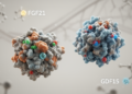
From an architectural standpoint, canonical bacterial enolase belongs to an ancient enzyme superfamily whose members stabilize an enolate intermediate through coordinated divalent metal ions, specifically magnesium, during the conversion of 2-phosphoglycerate into phosphoenolpyruvate. Structural characterization reveals that the polypeptide folds into a compact N-terminal alpha-beta domain alongside a larger C-terminal catalytic barrel. Although the active site and its flexible substrate-binding loops display rigorous evolutionary conservation across diverse bacterial phyla, peripheral loops, peptide termini, and oligomerization interfaces exhibit substantial sequence and conformational divergence. Monomers associate across a conserved dimer interface, and in कई bacterial lineages—including bacilli, streptococci, and staphylococci—enolase forms higher-order octamers assembled from four dimers. Crucially, the spatial organization of these oligomers dictates ligand accessibility. In Streptococcus pneumoniae, for example, the crystal structure demonstrates that C-terminal lysines are partially buried within the octameric interior, whereas an internal plasminogen-binding motif remains exposed on the solvent-accessible exterior surface. Consequently, linear sequence motifs alone cannot reliably predict interaction surfaces across living bacterial cells.
To evaluate these complex phenomena systematically, the review team implemented a structured, reproducible literature search of the PubMed and MEDLINE databases covering records from database inception through 27 October 2026. Applying an unrestricted search string yielded 607 candidate records, which were subsequently screened by title and abstract to exclude non-bacterial studies, generic citations lacking enolase-specific data, and redundant publications. From the 175 full-text articles assessed for detailed eligibility, 47 primary database reports fulfilled the inclusion criteria. Backward and forward citation searches identified an additional 35 qualifying investigations, establishing a core reference base of 82 primary research papers spanning structural biology, enzymology, RNA processing, membrane vesicle transport, host-pathogen interactions, and translational pharmacology. Because the assembled literature integrated disparate methodologies ranging from cryo-electron microscopy and recombinant binding assays to live-cell genetics and animal challenge models, the investigators synthesized the evidence through a critical narrative structure rather than an aggregated meta-analysis, noting that diverse mechanistic endpoints precluded a single statistical comparator.
Within the bacterial cytoplasm, enolase fulfills a well-documented moonlighting role by coupling metabolic status to post-transcriptional gene regulation through direct assembly into RNA-degradation machinery. In Escherichia coli, enolase serves as a stable component of the multi-enzyme RNA degradosome, joining polynucleotide phosphorylase and the DEAD-box helicase RhlB on the flexible non-catalytic scaffold of ribonuclease E. Structural studies confirm that a distinct segment of the ribonuclease E scaffold binds within a conserved cleft formed at the enolase dimer interface, physically uncoupling degradosome recruitment from secondary consequences of disrupted glycolysis. Functional analyses indicate that while enolase is dispensable for generic non-specific RNA turnover in reconstituted systems, its recruitment is required for the rapid degradation of specific transcripts during environmental perturbations. For instance, under phosphosugar stress, recruitment of enolase enables accelerated clearance of ptsG mRNA encoding the principal glucose permease, thereby arresting further sugar accumulation, while under oxygen deprivation, enolase-containing degradosomes modulate the stability of the small regulatory RNA DicF to alter cellular morphology.
Complementary intracellular functions have been characterized in Gram-positive bacteria, demonstrating that metabolic enzymes interact with distinct ribonucleolytic architectures across divergent evolutionary lineages. Gram-positive species lack an exact homolog of the ribonuclease E scaffold, yet organisms such as Bacillus subtilis assemble functional degradosome-like networks around ribonuclease Y, ribonuclease J enzymes, polynucleotide phosphorylase, and associated helicases. In Bacillus subtilis, enolase and phosphofructokinase copurify with this ribonuclease Y network. Biochemical evidence reveals that the small regulatory protein SR7P binds enolase directly, subsequently stimulating the ribonuclease activity of enolase-associated ribonuclease Y against specific cellular RNA substrates. The authors highlight this multi-protein assembly as evidence that enolase functions as a regulated interaction hub whose downstream effects are fine-tuned by auxiliary small proteins. Nonetheless, they caution that the physical recruitment of enolase in Gram-positive networks differs substantially from that observed in enteric bacteria, warning against assuming uniform mechanisms across disparate bacterial architectures where degradosome composition remains fluid and responsive to cellular physiology.
The detection of enolase outside the cell membrane introduces a fundamental topological paradox because canonical bacterial enolases entirely lack N-terminal Sec secretion signal peptides, hydrophobic transmembrane segments, and known covalent cell-wall anchoring motifs. Despite the absence of classical export signatures, intact-cell immunofluorescence, flow cytometry, immunoelectron microscopy, and surface proteomics have repeatedly detected enolase on the external envelope across Gram-positive and Gram-negative taxa. To explain this non-classical secretion, researchers have evaluated several non-exclusive mechanisms: direct membrane translocation via unknown channels, selective release coordinated with cell division or autolysis, packaging within extracellular membrane vesicles, and passive release from lysed cells followed by electrostatic surface reassociation. In Bacillus subtilis, enolase accumulates extracellularly during stationary phase without accompanying release of other cytosolic markers, a process disrupted by mutating a specific hydrophobic alpha-helix. Similarly, exponential-phase release of enolase in Staphylococcus aureus concentrates near division septa and correlates with major autolysin activity, suggesting that regulated peptidoglycan remodeling facilitates export without generalized cellular lysis.
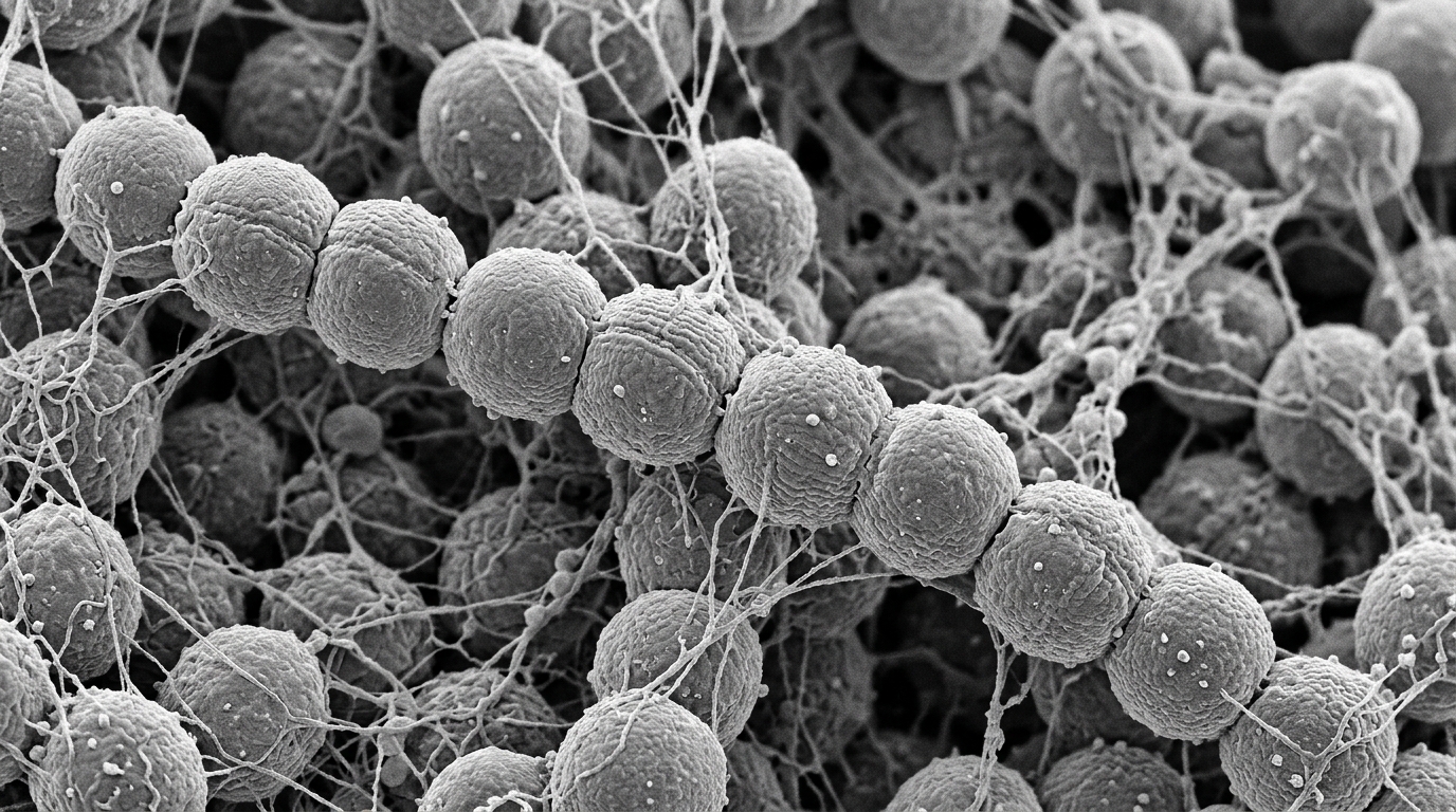
Extracellular vesicles represent an alternative vehicle for enolase transport, although cargo topology and physiological relevance vary markedly between bacterial lineages. Vesicular carriage has been described in organisms such as Bacteroides fragilis and group A streptococci. In group A streptococcal investigations, enolase remains protected against proteinase K digestion in intact extracellular vesicles and undergoes complete proteolysis only when vesicle membranes are disrupted with detergents, confirming that the enzyme resides within a membrane-delimited compartment rather than adhering as an external artifact. Furthermore, electrostatic reassociation accounts for substantial cell-surface localization in multiple Gram-positive species. In Lactobacillus crispatus, enolase and glyceraldehyde-3-phosphate dehydrogenase release into the medium under alkaline conditions and rebind to the cell surface at acidic pH through specific interactions with lipoteichoic acid. Similar reassociation events occur in Streptococcus pneumoniae and Leptospira interrogans, demonstrating that bacteria can capture extracellular enolase produced by neighboring cells, a phenomenon that complicates genetic knockouts and phenotypic interpretations in mixed bacterial communities.
On the external cell surface, the dominant host-interaction paradigm involves the recruitment of human plasminogen, the circulating zymogen of the fibrinolytic serine protease plasmin. By immobilizing plasminogen on the bacterial surface, pathogens can exploit host tissue-type or urokinase-type plasminogen activators to generate active plasmin, acquiring broad surface-associated proteolytic activity that degrades fibrin clots, laminin, and extracellular matrix barriers. In Streptococcus pneumoniae, an internal nine-residue exposed loop comprising the sequence FYDKERKVY mediates plasminogen binding, and site-directed mutagenesis of this region diminishes adherence and virulence in intranasal murine challenge models. However, this motif is not universal; Lactobacillus plantarum relies on both internal and C-terminal basic lysine residues, while oral streptococci activate plasminogen despite significant sequence divergence across homologous segments. Beyond plasminogen capture, surface-associated enolases from pathogens and commensals alike bind extracellular matrix components—such as fibronectin, laminin, collagen, and fibrinogen—and engage human complement regulators, including C4b-binding protein and factor H, to attenuate opsonophagocytosis and immune clearance.
To overcome recurrent methodological pitfalls in the moonlighting literature, the review establishes a rigorous seven-criterion evaluation framework that replaces former linear evidence hierarchies. This heuristic framework grades evidence across seven complementary operational tiers: in silico sequence or structural prediction, extracellular or envelope-enriched proteomic detection, validated intact-cell compartmental localization with membrane-integrity controls, quantitative biochemical binding demonstrating concentration dependence and partner specificity, relevant cellular phenotypes modulated by competitive blocking, targeted separation-of-function mutagenesis preserving catalytic flux and cellular growth, and validation within physiologically compatible host systems. The authors illustrate the necessity of this framework by noting that many published claims rely solely on recombinant protein binding or unverified deletions that impair central metabolism. For example, in Streptococcus suis, the eno251 substitution preserved overall bacterial growth while impairing endothelial monolayer translocation, yet subsequent kinetic testing demonstrated altered substrate affinity, illustrating that preserved growth alone does not prove complete metabolic neutrality or an exclusively non-catalytic phenotype.
Translational initiatives targeting bacterial enolase must carefully negotiate substantial biochemical hurdles, structural conservation, and functional redundancy. Developing active-site inhibitors faces the primary obstacle of high sequence identity with human enolase isoforms ENO1, ENO2, and ENO3, raising risks of host toxicity. Although natural phosphonates such as SF2312 exhibit nanomolar potency against purified Escherichia coli enolase, their charged transition-state structures exhibit poor membrane permeability, and efficacy varies dramatically with available carbon sources. Conversely, targeting exposed host-binding interfaces offers potential pathogen selectivity without disrupting host metabolism, yet functional redundancy among multiple bacterial adhesins and plasminogen receptors frequently compensates for single-target inhibition. Similarly, while surface enolase serves as a protective subunit vaccine candidate in several animal challenge systems—including models of Streptococcus iniae, Streptococcus suis, and Mycoplasmopsis synoviae—other controlled trials have documented high antibody titers without protection against lethal challenge. The authors conclude that future clinical translation requires rigorous separation-of-function genetics, quantitative localization standards, and head-to-head evaluation in natural-host infection models.
Subject of Research: Bacterial enolase localization and moonlighting functions in cellular physiology and pathogenesis
Article Title: Beyond glycolysis: bacterial enolase as a localization-dependent moonlighting protein in physiology and pathogenesis
Article References: Lu, F., Ding, M., Liu, Z., Peng, J., & Zheng, K. (2026). Beyond glycolysis: bacterial enolase as a localization-dependent moonlighting protein in physiology and pathogenesis. Frontiers in Microbiology, 17, Article 1978482. https://doi.org/10.3389/fmicb.2026.1978482
Image Credits: AI Generated
DOI: 10.3389/fmicb.2026.1978482
Keywords: bacterial enolase, protein moonlighting, glycolytic enzymes, RNA degradosome, plasminogen recruitment, non-classical secretion, extracellular vesicles, cell envelope reassociation, RNase E, ribonuclease Y, Streptococcus pneumoniae, Staphylococcus aureus, separation-of-function genetics, complement evasion, biofilm matrix, antimicrobial targets, active-site inhibition, subunit vaccines
News Source: Drew Townsend. (November 17, 2026). Bacterial Enolase Moonlighting Functions Depend on Cellular Localization and Environmental Conditions. Scienmag.