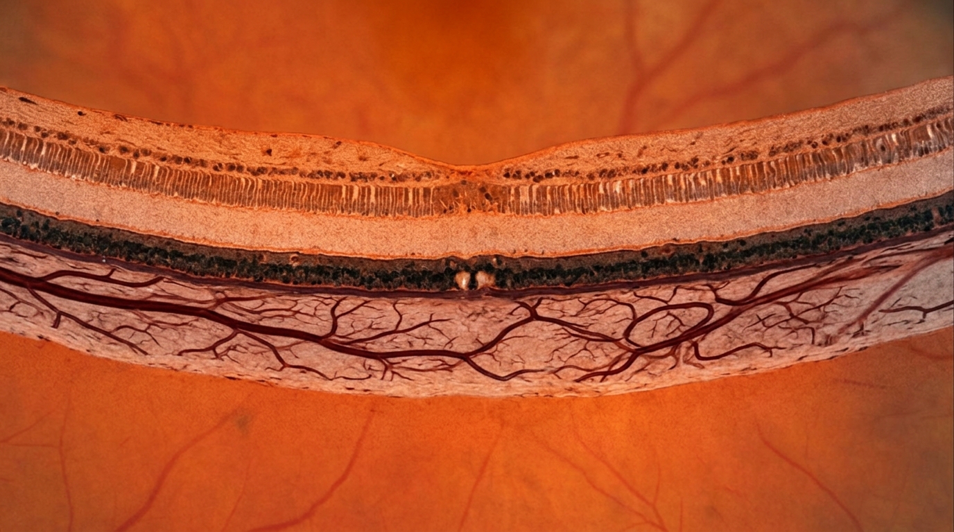
Early changes in choroidal vascular structure can predict long-term refractive progression in children receiving low-dose atropine, yet this predictive capacity does not automatically identify the specific therapeutic benefit attributable to the medication itself. In an analysis examining recent findings from the AMIXT randomized trial, researcher Jing Chen evaluated how four-month alterations in perifoveal luminal volume correlate with two-year refractive outcomes. The original investigation by Zuo and colleagues demonstrated that early three-dimensional choroidal vascular remodeling serves as a measurable indicator of subsequent myopic progression during daily treatment with 0.01 percent atropine. While these vascular metrics provide valuable clinical insights, interpreting what these structural shifts mean requires distinguishing between a patient’s natural ocular growth trajectory and their genuine biological response to pharmacologic intervention.
The investigated clinical cohort comprised children diagnosed with both myopia and intermittent exotropia who underwent three-dimensional choroidal imaging during the AMIXT clinical trial. By tracking volumetric adjustments in the choroid over time, researchers identified four-month changes in perifoveal luminal volume, designated as ΔLV₃, as a meaningful predictor of refractive progression at twenty-four months. Alongside these imaging observations, accompanying experimental studies revealed that choroidal endothelial oxidative stress and nitric oxide signaling pathways participate in this process. These laboratory findings offer biological plausibility for the role of vascular tissue in eye growth regulation, suggesting that the vascular layer actively responds during myopia management rather than acting as a passive tissue layer.
Despite the predictive accuracy of the ΔLV₃ vascular metric, prediction models categorize clinical outcomes based on observed rates of refractive change rather than comparative drug efficacy. Children who display slower refractive progression while using 0.01 percent atropine might possess an inherently favorable growth trajectory, a robust physiological response to the medication, or a combination of both factors. Conversely, continued refractive progression in a treated child does not inherently demonstrate a failure of drug benefit, as that individual might have experienced substantially faster myopic worsening without any therapeutic intervention. Consequently, an early choroidal metric may successfully pinpoint individuals who remain at risk of rapid progression without identifying which patients derive the least pharmacological benefit from atropine.
Broader randomized clinical trial data provide essential context for understanding how choroidal thickness changes relate to both natural ocular expansion and pharmacological responses. In the MOSAIC trial, greater choroidal thickening was associated with reduced axial elongation and slower refractive progression among children in both the atropine-treated groups and the placebo control groups. Furthermore, formal mediation analyses within that trial indicated that the overall effect of atropine on choroidal thickening was only partly explained by alterations in axial length or refractive error. These comparative patterns confirm that choroidal modifications capture dual signals, reflecting both the general trajectory of childhood eye enlargement and medication-specific biological adaptations.
Additional evidence from the LAMP trial reinforces this complex relationship across varying drug concentrations. Investigators in that trial documented concentration-dependent choroidal thickening that accompanied slower overall myopia progression, with statistical mediation analysis supporting a partial contribution of choroidal changes to the overall treatment effect observed at 0.05 percent atropine. Although two-dimensional measurements of choroidal thickness are not directly interchangeable with volumetric three-dimensional luminal volume assessments, both lines of investigation demonstrate that choroidal remodeling reflects both drug-induced responses and underlying developmental eye growth. These shared observations highlight the need for careful statistical and physiological differentiation when evaluating choroidal imaging biomarkers in pediatric populations.
Mediation analyses in the study by Zuo and collaborators identified statistically significant indirect effects operating through twenty-four-month choroidal volume changes, while cellular assays demonstrated the involvement of endothelial oxidative stress and nitric oxide pathways. Together, these complementary findings support the concept that the choroid serves as an active cellular participant during atropine-mediated myopia control. However, establishing biological plausibility and long-term volume mediation does not resolve the clinical interpretation of the earlier four-month ΔLV₃ measurement. Demonstrating that an early metric predicts subsequent refractive progression during an active treatment regimen does not prove that it can distinguish variations in the true treatment benefit generated by atropine.
For translation into routine ophthalmic practice, perifoveal luminal volume measurements may eventually help clinicians identify pediatric patients at elevated risk of continuing refractive progression, provided the predictive validity of ΔLV₃ receives independent clinical confirmation. Clinical specialists must exercise caution, as a small early increase in luminal volume should not automatically be interpreted as medication failure, especially when axial elongation and refractive progression remain the definitive outcomes of clinical concern. Moving forward, clinical investigations must evaluate whether early choroidal monitoring can differentiate between children who should remain on 0.01 percent atropine and those who require higher drug concentrations or supplementary interventions to achieve effective myopia control.
Subject of Research: Choroidal vascular remodeling and outcome prediction in childhood myopia atropine therapy
Article Title: From predicting myopia progression to identifying treatment benefit: interpreting choroidal remodeling during atropine therapy
Article References: Chen, J. (2026). From predicting myopia progression to identifying treatment benefit: interpreting choroidal remodeling during atropine therapy. Journal of Translational Medicine, 24(1), Article 1430. https://doi.org/10.1186/s12967-026-09224-5
Image Credits: AI Generated
DOI: 10.1186/s12967-026-09224-5
Keywords: myopia progression, atropine therapy, choroidal remodeling, luminal volume, perifoveal luminal volume, AMIXT trial, MOSAIC trial, LAMP trial, intermittent exotropia, refractive progression, axial elongation, choroidal thickness, endothelial oxidative stress, nitric oxide signaling, mediation analysis, pediatric ophthalmology
News Source: Ophelia Keating. (November 17, 2026). Early Choroidal Changes Predict Myopia Progression but May Not Show Treatment Benefit. Scienmag.