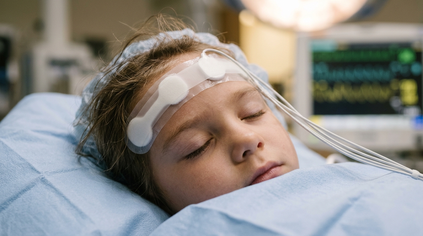
Children with functional single-ventricle anatomy demonstrate distinct frontal brainwave signatures during surgical anesthesia compared to peers with two working pumping chambers, according to a prospective observational study published in BMC Anesthesiology. Anesthesiology researchers identified that pediatric patients undergoing the Fontan operation display a significant shift toward slower cerebral oscillations alongside marked reductions in high-frequency electrical activity during midazolam-maintained sedation.
Intraoperative electroencephalographic monitoring and precise anesthetic titration represent critical clinical priorities for children with functional single-ventricle physiology who present for staged cardiac palliation. Despite the recognized clinical importance of maintaining tailored anesthetic depth in this vulnerable surgical cohort, the baseline electroencephalographic characteristics that define a stable anesthetic state in these young patients had previously remained undefined against comparative surgical controls.
To investigate these neurophysiological differences, the prospective study evaluated forty young children undergoing cardiac surgery, divided evenly into two distinct physiological cohorts. The single-ventricle group comprised twenty children who had previously received bidirectional Glenn palliation and were currently presenting for a completion Fontan procedure. For comparison, investigators enrolled twenty age-matched control subjects aged two to six years who possessed biventricular physiology and were undergoing surgical repair for acyanotic congenital heart defects.
Investigators monitored neural activity using a four-channel frontal electroencephalogram during an established, stable anesthetic state induced and maintained with the sedative midazolam. The research team analyzed comprehensive spectral parameters across the monitoring period, evaluating total spectral power, band-specific absolute and relative power measurements, combined low-frequency power spanning the delta and theta bands, high-frequency activity across the alpha, beta, and gamma spectra, and the computed theta-to-beta ratio.
The spectral analyses uncovered pronounced neurophysiological divergences between the two surgical cohorts. Children with functional single-ventricle anatomy exhibited significantly reduced absolute gamma power at 11.7 decibels compared with 13.4 decibels in the biventricular control group, reflecting a statistically significant mean difference with a ninety-five percent confidence interval spanning minus 4.1 to minus 0.4 decibels. Concurrently, the single-ventricle cohort demonstrated a clear shift toward lower frequencies, recording significantly higher relative theta power at 7.2 percent versus 3.8 percent in controls, alongside elevated total low-frequency power measuring 29.3 percent compared with 19.3 percent in the biventricular group.
These spectral shifts were further emphasized by a significantly higher theta-to-beta ratio among single-ventricle patients, averaging 0.23 compared to 0.09 in biventricular controls. To account for potential baseline neurological confounding, the researchers performed planned subgroup analyses restricted exclusively to participants with normal developmental status, defined as scores greater than or equal to minus one standard deviation after excluding children with documented developmental delays. Even within this developmentally normal subgroup, patients with single-ventricle anatomy continued to demonstrate significantly higher relative low-frequency power and diminished high-frequency power compared with their biventricular peers.
The authors concluded that children presenting with functional single-ventricle physiology manifest unique anesthesia-related frontal electroencephalographic signatures governed by slower oscillatory rhythms and attenuated high-frequency spectral components. The team noted that these characteristic patterns suggest potentially altered cortical responsiveness to midazolam anesthesia in this patient population relative to children with biventricular anatomy, underscoring the distinct neurophysiological substrate present during complex congenital heart surgery.
Subject of Research: Intraoperative frontal electroencephalographic patterns in children with single-ventricle versus biventricular physiology
Article Title: Distinct intraoperative electroencephalographic features in children with single-ventricle versus biventricular physiology
Article References: Jang, H.-Y., Song, I.-K., Joe, Y.-E., Kim, S.-H., Bang, J.-Y., & Shin, W.-J. (2026). Distinct intraoperative electroencephalographic features in children with single-ventricle versus biventricular physiology. BMC Anesthesiology. https://doi.org/10.1186/s12871-026-04368-9
Image Credits: AI Generated
DOI: 10.1186/s12871-026-04368-9
Keywords: electroencephalography, intraoperative EEG, single-ventricle physiology, Fontan procedure, biventricular physiology, bidirectional Glenn palliation, midazolam, acyanotic congenital heart defects, frontal EEG, gamma power, theta power, low-frequency power, theta/beta ratio, pediatric cardiac surgery, cortical responsiveness, developmental status, spectral power analysis
News Source: Harold Sullivan. (November 17, 2026). Single Ventricle Physiology Alters Frontal Brainwave Patterns During Pediatric Anesthesia. Scienmag.