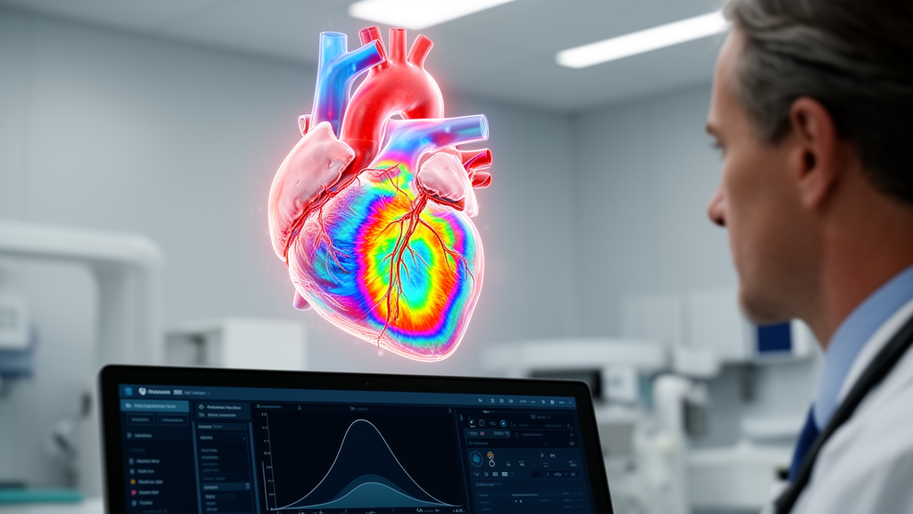
The intraoperative Focused 3D Method enabled the accurate identification of the location and mechanism of residual mitral regurgitation in two male patients undergoing robot-assisted mitral valve repair, according to a case report published in JA Clinical Reports. The imaging technique provided critical anatomical details during surgery that directly informed clinical decision-making regarding reintervention.
Rapid and precise intraoperative evaluation of residual regurgitation is considered essential following robot-assisted repairs of the mitral valve. However, pinpointing the exact underlying etiology of residual leakage using conventional two-dimensional transesophageal echocardiography, known as 2D-TEE, can present significant clinical challenges for surgical and echocardiographic teams.
In the first reported case, findings obtained with the Focused 3D Method revealed a previously unrecognized A1 prolapse following the initial repair for severe mitral regurgitation. This intraoperative observation prompted the surgical team to initiate a second pump run to successfully correct the leaflet defect. In the second patient, the imaging method clearly demonstrated shallow leaflet coaptation, providing evidence that supported the team’s decision to avoid an unnecessary surgical reintervention.
Both patients experienced favorable postoperative outcomes following their robotic procedures. Based on these two cases, the researchers concluded that the Focused 3D Method may enhance intraoperative assessment of residual mitral regurgitation and assist surgical teams in determining whether a second pump run is warranted after mitral valve repair.
Subject of Research: Intraoperative assessment of residual mitral regurgitation using the Focused 3D Method
Article Title: Evaluation of residual mitral regurgitation after robot-assisted mitral valve repair using the focused 3D method: a report of two cases
Article References: Miyata, K., Shigematsu, S., Sato, M., & Okita, A. (2026). Evaluation of residual mitral regurgitation after robot-assisted mitral valve repair using the focused 3D method: a report of two cases. JA Clinical Reports. https://doi.org/10.1186/s40981-026-00890-4
Image Credits: AI Generated
DOI: 10.1186/s40981-026-00890-4
Keywords: Focused 3D Method, residual mitral regurgitation, robot-assisted mitral valve repair, mitral regurgitation, mitral valve repair, transesophageal echocardiography, 2D-TEE, intraoperative assessment, A1 prolapse, leaflet coaptation, second pump run, surgical reintervention, case report, postoperative outcomes, JA Clinical Reports, Kazuto Miyata
News Source: Ophelia Keating. (November 16, 2026). Focused 3D Method Aids Residual Mitral Regurgitation Assessment in Two Cases. Scienmag.