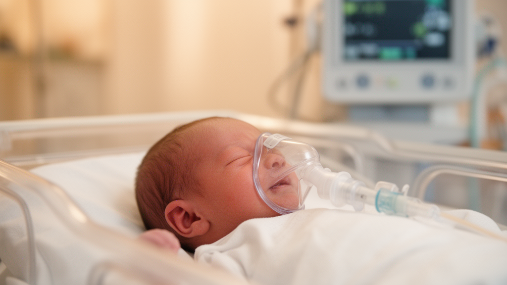
In clinically stable very preterm infants, transitioning from standard continuous positive airway pressure to non-invasive intermittent positive pressure ventilation maintains stable cardiac output while substantially decreasing diaphragmatic effort and enhancing pulmonary aeration, according to a randomized crossover trial conducted at Siriraj Hospital in Bangkok, Thailand. The clinical investigation systematically compared the immediate hemodynamic and respiratory impacts of three distinct non-invasive ventilation strategies, specifically addressing longstanding concerns that the delivery of higher mean airway pressures to fragile neonatal lungs might inadvertently impede venous return or compromise biventricular performance.
Non-invasive respiratory support serves as an essential standard of care in neonatal intensive care units to recruit collapsed alveoli, expand functional residual capacity, improve oxygenation, and minimize the risk of bronchopulmonary dysplasia and ventilator-associated lung injury by reducing the necessity for invasive endotracheal intubation. While nasal continuous positive airway pressure (NCPAP) remains the conventional primary modality for preterm infants experiencing acute respiratory distress, nasal intermittent positive pressure ventilation (NIPPV) provides supplementary inflations above baseline positive end-expiratory pressure. These intermittent inflations increase mean airway pressure and facilitate carbon dioxide clearance, yet clinicians have frequently questioned whether elevated intrathoracic pressures might impair right or left ventricular filling and cardiac output.
To evaluate these cardiopulmonary responses, investigators designed a prospective, randomized, two-sequence crossover study involving preterm neonates born at less than 32 weeks of gestation or with a birth weight below 1500 grams who were already receiving stable NCPAP support. The trial evaluated thirty infants enrolled between postnatal days 3 and 14, an enrollment window specifically chosen to minimize the physiological confounding associated with early transitional circulatory adjustments while avoiding the confounding effects of advanced, evolving chronic lung disease. Infants with major congenital anomalies, critical congenital heart disease other than patent ductus arteriosus or atrial septal defects, or congenital brain malformations were systematically excluded.
The study protocol exposed each participant to four consecutive two-hour epochs, beginning with a baseline NCPAP period of at least two hours to confirm baseline stability, followed by randomization to either non-synchronized NIPPV (NS-NIPPV) or neurally synchronized NIPPV (S-NIPPV). Following the first intervention phase, infants underwent a two-hour washout period on baseline NCPAP before crossing over to the alternate NIPPV mode for an additional two hours. All ventilatory modes were administered via a Servo-N ventilator, with neural synchronization achieved through non-invasive neurally adjusted ventilatory assist (NIV-NAVA) guided by an inserted diaphragmatic electrical activity (Edi) catheter. Ventilator settings across modalities were carefully titrated by bedside clinical staff to maintain mean airway pressure within plus or minus 2 cmH2O of baseline NCPAP levels.
Rigorous blinding was maintained throughout the evaluation periods; outcome assessors and the offline image analyst remained blinded to the ventilation mode, and the ventilator display screen was completely covered with an opaque shield. During the final thirty minutes of each two-hour epoch, a neonatology fellow with specialized echocardiographic and sonographic training acquired functional echocardiograms and twelve-zone lung ultrasound examinations. To ensure measurement precision, all outflow tract diameters and velocity-time integrals were subsequently re-verified by a second blinded investigator, demonstrating strong inter-observer agreement with intraclass correlation coefficients ranging from 0.75 to 0.90 across evaluated parameters.
Primary hemodynamic outcomes centered on right ventricular output (RVO) and left ventricular output (LVO), calculated through mid-systolic valve annulus diameters, Doppler velocity-time integrals, and heart rates. Across all four time epochs, linear mixed-effects modeling revealed no significant carryover, period, or sequence effects, confirming the statistical integrity of the crossover design. The analysis demonstrated that biventricular output remained well preserved across all three non-invasive support modes, with no significant differences observed in right or left ventricular output between NCPAP, NS-NIPPV, and S-NIPPV.
Right ventricular output consistently exceeded left ventricular output across all study modes, maintaining an average RVO to LVO ratio of approximately 2.1, a finding consistent with the 60 percent prevalence of interatrial left-to-right communication within the cohort, as well as a larger pulmonary annulus diameter and higher pulmonary velocity-time integral. When comparing the two intermittent modes, S-NIPPV demonstrated a non-significant numerical trend toward higher right ventricular output compared to NS-NIPPV, exhibiting a mean difference of 24 mL/kg/min (95% CI: -3 to 51; p = 0.08), whereas no meaningful difference emerged between S-NIPPV and baseline NCPAP.
Secondary evaluations of ventricular systolic performance and myocardial deformation utilizing speckle-tracking echocardiography provided detailed insights into cardiac mechanics. Right ventricular functional indices, including tricuspid annular plane systolic excursion, functional area change, and tissue Doppler velocities, were comparable across ventilation strategies. However, left ventricular peak global longitudinal strain and strain rate demonstrated significantly greater negative magnitude during NS-NIPPV compared to NCPAP (-2.54% [95% CI: -4.61 to -0.48; p = 0.02] and -0.45 [95% CI: -0.78 to -0.11; p = 0.008], respectively), while S-NIPPV showed higher shortening fraction and ejection fraction by M-mode compared with NS-NIPPV.
Continuous monitoring of respiratory mechanics revealed distinct advantages for synchronized pressure delivery. Peak electrical activity of the diaphragm, an objective measure of diaphragmatic workload and respiratory muscle effort, was significantly reduced during S-NIPPV (7.2 ± 5.1 µV) compared with baseline NCPAP (10.0 ± 6.8 µV; p < 0.01) and NS-NIPPV (9.3 ± 6.5 µV). Simultaneously, mean airway pressure was modestly higher during S-NIPPV (6.7 ± 1.0 cmH2O) and NS-NIPPV (6.6 ± 0.7 cmH2O) than during NCPAP (5.8 ± 0.7 cmH2O; p < 0.001), reflecting the active intermittent inflations delivered above positive end-expiratory pressure.
Parallel lung ultrasound assessments confirmed significant improvements in pulmonary aeration when active pressure support was delivered. Total lung ultrasound scores, scored across twelve distinct anatomical zones, decreased significantly during both NS-NIPPV (mean difference -2.52; 95% CI: -3.64 to -1.39; p < 0.001) and S-NIPPV (mean difference -2.22; 95% CI: -3.34 to -1.10; p < 0.001) relative to NCPAP. These aeration gains were particularly pronounced in the dependent posterior lung fields, with no significant difference detected between the two NIPPV modalities themselves.
The study authors noted several methodological considerations, observing that the cohort consisted predominantly of clinically stable preterm infants, which limits the direct extrapolation of these physiological findings to critically ill, extremely unstable neonates receiving inotropic support. Furthermore, because secondary echocardiographic and strain analyses involved numerous pairwise comparisons without formal multiplicity adjustments, isolated functional differences must be interpreted as hypothesis-generating. The investigators concluded that short-term non-invasive ventilation via S-NIPPV and NS-NIPPV successfully enhances pulmonary recruitment and decreases breathing effort without exerting adverse hemodynamic penalties on neonatal biventricular performance.
Detailed participant flow and baseline clinical profiles offer essential context for the trial findings. Out of fifty-six infants initially assessed for eligibility, twenty-six were excluded, leaving thirty enrolled neonates. One infant assigned to receive non-synchronized ventilation first was administered neurally synchronized ventilation due to a technical error, resulting in fourteen infants analyzed in the non-synchronized sequence and sixteen in the synchronized sequence for the primary as-received analysis, while an intention-to-treat analysis of fifteen infants per group yielded comparable ventricular outputs. Across the cohort, mean gestational age was 30.0 ± 2.2 weeks in the non-synchronized sequence and 29.9 ± 1.9 weeks in the synchronized sequence, with enrollment at an average of 6 ± 2 and 8 ± 3 days after birth. Although birthweight and surfactant exposure were similar between groups, infants randomized to synchronized support first had slightly higher baseline positive end-expiratory pressure (6.3 ± 0.7 versus 5.8 ± 0.4 cmH2O; p = 0.04) and more frequent prior invasive mechanical ventilation (44 percent versus 7 percent; p = 0.04).
Beyond primary ventricular outputs, secondary echocardiographic metrics revealed subtle, mode-specific variations in cardiac mechanics. While conventional right ventricular performance markers such as tricuspid annular excursion and fractional area change did not differ, non-synchronized NIPPV was associated with a statistically significant increase in right ventricular tissue Doppler a-prime velocity compared with NCPAP (mean difference 0.90 cm/s; 95% CI: 0.27 to 1.53; p = 0.008). On left ventricular assessment, systolic performance evaluated by the five-sixths area-length method was higher during non-synchronized NIPPV than during baseline continuous pressure (mean difference 3.20; 95% CI: 0.28 to 6.13; p = 0.03), and neurally synchronized NIPPV demonstrated higher shortening fraction (mean difference 3.35; 95% CI: 0.83 to 5.86; p = 0.01) and ejection fraction (mean difference 4.76; 95% CI: 1.04 to 8.48; p = 0.01) relative to non-synchronized support. Radial and circumferential myocardial strain parameters exhibited no significant shifts across modalities, and global myocardial performance indices remained stable, suggesting that observed variations were modest in magnitude.
These cardiopulmonary findings also provide mechanistic context when interpreted alongside earlier neonatal literature. Previous investigations examining respiratory support have reported conflicting hemodynamics, with some studies observing reduced right ventricular output and venous return at modest pressure levels, whereas others noted preserved output at higher distending pressures. The research team hypothesized that the attenuated left ventricular longitudinal strain seen on unassisted continuous pressure may stem from unassisted work of breathing, which transiently increases cardiac afterload and myocardial wall stress. The investigators highlighted several physiological boundaries of their study, noting that transpulmonary pressure, carbon dioxide concentrations, and specific left-to-right shunt volumes across atrial or ductal communications were not quantified. Furthermore, because each ventilatory epoch was restricted to two hours, the authors emphasized that their exploratory findings reflect only immediate physiological responses, leaving the long-term cardiopulmonary outcomes of synchronized and non-synchronized non-invasive strategies to be defined in larger multicenter trials.
Subject of Research: Hemodynamic and cardiopulmonary effects of non-invasive ventilation modes in preterm infants
Article Title: Hemodynamic and respiratory effects of non-invasive ventilation modalities in very preterm infants: a randomized crossover trial
Article References: Naknin, N., Yangthara, B., Kitsommart, R., Altit, G., & Wutthigate, P. (2026). Hemodynamic and respiratory effects of non-invasive ventilation modalities in very preterm infants: a randomized crossover trial. Pediatric Research. https://doi.org/10.1038/s41390-026-05593-1
Image Credits: AI Generated
DOI: 10.1038/s41390-026-05593-1
Keywords: preterm infants, non-invasive ventilation, nasal continuous positive airway pressure, synchronized NIPPV, non-synchronized NIPPV, neurally adjusted ventilatory assist, electrical activity of the diaphragm, ventricular output, echocardiography, speckle-tracking echocardiography, lung ultrasound score, myocardial performance, global longitudinal strain, neonatal intensive care, pulmonary aeration, crossover trial
News Source: Harold Sullivan. (November 14, 2026). Synchronized non-invasive ventilation preserves preterm cardiac output while easing breathing effort. Scienmag.