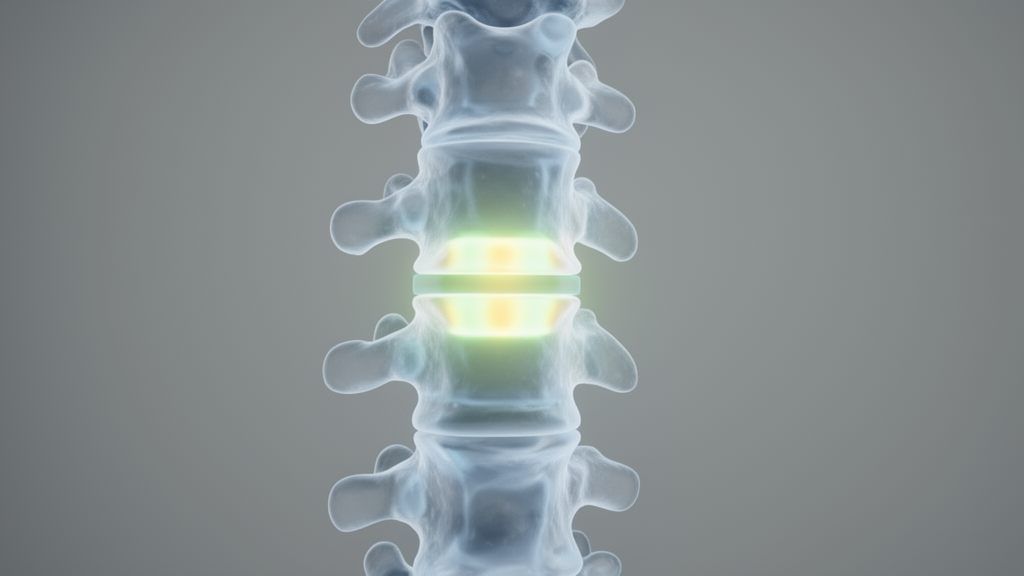
Researchers from Fujian Provincial Hospital in China have demonstrated that co-modifying bone marrow mesenchymal stem cells (BMSCs) with two distinct growth factors significantly enhances spinal fusion in a rat model. Published in Frontiers in Cell and Developmental Biology, the study reveals that while single-gene modifications showed limited efficacy, the simultaneous expression of fibroblast growth factor 1 (FGF1) and bone morphogenetic protein 2 (BMP2) resulted in an 80% fusion rate, compared to 0% for controls and FGF1-only groups. This finding suggests a synergistic mechanism where the two proteins work together to promote robust bone formation, offering a potential new direction for treating spinal disorders where achieving stable bone fusion remains a clinical challenge.
The study, led by Wubing He and colleagues, focused on the limitations of current BMSC-based therapies. Although BMSCs are multipotent cells capable of differentiating into osteoblasts, chondrocytes, and adipocytes, their ability to induce robust bone formation in vivo has been inconsistent. The research team aimed to determine whether combining FGF1, known for promoting cell proliferation and angiogenesis, with BMP2, a potent osteoinductive factor, could overcome these limitations. By using lentiviral vectors to introduce both genes into the stem cells, the investigators sought to create a cellular therapy that could more effectively bridge spinal defects.
To test this hypothesis, the researchers isolated BMSCs from three-week-old male Sprague-Dawley rats. These cells were transduced with lentiviruses carrying FGF1, BMP2, or both genes, alongside a control group receiving an empty vector. In vitro assessments confirmed that the dual-modified cells exhibited significantly higher transgene expression, proliferation rates, and alkaline phosphatase (ALP) activity compared to single-gene or control groups. The dual-modified cells also showed upregulated expression of osteogenic markers such as Runx2 and OPG, indicating a stronger propensity for bone differentiation.
Transcriptomic analysis provided further insight into the molecular mechanisms driving these effects. RNA sequencing of the dual-modified cells identified 2,313 differentially expressed genes, including 1,257 upregulated and 1,056 downregulated genes. The upregulated genes were primarily associated with cell proliferation, angiogenesis, and ossification, while inflammatory genes were suppressed. Pathway enrichment analysis highlighted the activation of the PI3K-Akt and MAPK signaling pathways, suggesting that FGF1 and BMP2 may act synergistically through these intracellular cascades to enhance bone regeneration.
The in vivo component of the study involved implanting the modified cells into the lumbar vertebrae of 52 seven-week-old male rats. The animals were randomly allocated into four groups: LV-Vector, LV-FGF1, LV-BMP2, and LV-FGF1-BMP2. Each group received 5 × 10^6 cells loaded onto absorbable collagen sponges. The surgical procedure involved exposing the lumbar 4–5 vertebrae and implanting the cell-laden sponges on both sides of the vertebrae. The study was conducted in compliance with ARRIVE 2.0 guidelines, and all animal procedures were approved by the Institutional Animal Care and Use Committee of Fujian Provincial Hospital.
Biosafety assessments indicated that the local expression of FGF1 and BMP2 did not lead to significant systemic elevation of these proteins. Serum levels measured by ELISA at one, two, and three weeks post-operation showed no significant differences among the groups. Furthermore, histological examination of major organs, including the heart, liver, lung, and kidney, revealed no obvious abnormalities or toxic effects.
At the eight-week endpoint, the primary outcome was assessed through manual palpation, micro-CT imaging, and histological analysis. Manual palpation, performed by two independent observers blinded to group allocation, revealed a stark contrast in fusion rates. The LV-FGF1-BMP2 group achieved an 80% fusion rate (8 out of 10 rats), which was significantly higher than the 20% rate observed in the LV-BMP2 group (2 out of 10). In contrast, both the LV-Vector and LV-FGF1 groups showed a 0% fusion rate. Statistical analysis using Fisher’s exact test confirmed that the fusion rate in the dual-gene group was significantly higher than in the other groups, with a p-value of 0.001 compared to the vector control.
Micro-CT imaging corroborated the palpation findings, showing marked differences in callus formation. The LV-FGF1-BMP2 group exhibited the most prominent callus formation with complete laminar fusion, whereas the LV-BMP2 group showed significant callus but incomplete fusion. The LV-FGF1 and LV-Vector groups displayed minimal or no significant callus formation. Histological analysis using H&E and Masson’s trichrome staining further supported these results, demonstrating substantial new bone formation in the dual-gene group that met fusion criteria, while other groups showed limited or no bridging bone.
Immunohistochemical staining of the fusion sites revealed higher expression of osteogenic markers Runx2 and Osterix, as well as angiogenic markers CD31 and VEGFA, in the LV-FGF1-BMP2 group. This indicates that the dual modification not only promoted bone formation but also enhanced the vascularization of the fusion site, which is critical for sustaining new bone tissue. The combination of these markers suggests a more favorable microenvironment for bone healing, where angiogenesis and osteogenesis are tightly coupled.
Despite the promising results, the authors acknowledge several limitations. The study was conducted in a rat model, which may not fully translate to human clinical applications. The sample size for the confirmatory endpoints was relatively small, with n = 10 per group, which limits the precision of the estimates. Additionally, the biosafety assessment was limited to serum levels and one-week organ histology, which may not capture long-term risks such as tumorigenicity or abnormal bone formation. The authors emphasize that further validation in larger animal models and human clinical trials is necessary to confirm the safety and efficacy of this approach.
The findings highlight the potential of dual-gene modification as a strategy to enhance the osteogenic potential of BMSCs. By leveraging the complementary roles of FGF1 and BMP2, the researchers were able to achieve a higher fusion rate than previously reported with single-gene approaches. This approach could offer a new perspective for spinal fusion therapy, potentially reducing recovery time and improving surgical outcomes. However, the precise molecular mechanisms underlying the synergy between FGF1 and BMP2 remain to be fully elucidated, and future studies should focus on optimizing gene delivery and expression timing.
In conclusion, this study provides experimental evidence that co-modification of BMSCs with FGF1 and BMP2 exerts synergistic effects on osteogenesis and spinal fusion. The dual-gene approach significantly enhanced cell proliferation and osteogenic differentiation in vitro and achieved a high fusion rate in vivo. While further research is needed to address safety and efficacy in larger models, these results represent a significant step forward in the development of gene-modified stem cell therapies for spinal disorders.
Beyond baseline osteogenic markers, the transcriptomic profiling identified specific molecular drivers within the enriched signaling cascades. Differential expression analysis revealed marked upregulation of critical proliferation genes, such as Ccnd1, E2f1, Cdk1, Ccnb1, and Areg, alongside ossification regulators including Wnt5a, Tgfb1, Spp1, Igfbp5, and Cyp26b1. Angiogenic mediators, including Flt1, Hgf, Notch4, Mmp9, Serpine1, and Ccbe1, were also elevated, whereas inflammatory genes such as Casp4, Lbp, Vcam1, C4a, C6, and Aoc3 were significantly downregulated. Protein-protein interaction network analyses further highlighted central hub genes connecting these programs. Within the upregulated PI3K-Akt cascade, Il6, Csf1r, Csf3, Tlr4, and Flt1 served as core hubs, while the MAPK pathway was centered around Il1b, Csf1r, Flt1, Hgf, and Tgfb1. The investigators noted that directly verifying these causal mechanisms will require dedicated pharmacological inhibition experiments targeting PI3K/Akt or MEK/ERK alongside phosphorylated protein measurements.
The surgical evaluation also yielded detailed statistical comparisons defining the relative advantages of the dual-gene intervention over single-factor and control approaches. When evaluating manual palpation outcomes, animals in the control and FGF1-only groups faced a fivefold greater risk of non-fusion compared to the dual-gene cohort (relative risk 5.00, 95% confidence interval 1.448 to 17.271). In addition, the 80% fusion rate achieved by the dual-gene group was four times that of the BMP2 single-gene group (relative risk 4.00, 95% confidence interval 1.115 to 14.354; p = 0.023), corresponding to a fusion odds ratio of 16.00 (95% confidence interval 1.788 to 143.150). Prior to implantation, the phenotypic identity of the harvested stem cells was rigorously verified through flow cytometry, confirming positive surface expression of CD29 and CD90 coupled with negative expression of hematopoietic markers CD34 and CD45, while functional multipotency was evidenced by Alizarin Red mineralization and Oil Red O lipid staining.
Methodological constraints associated with the rat posterolateral fusion model also defined the interpretive boundaries of the study. Because of technical limitations inherent to the small-animal model, the investigators did not perform quantitative micro-CT density assessments or formal quantitative histomorphometric measurements, leaving structural bone parameters incompletely characterized. Furthermore, the biomechanical strength and long-term load-bearing properties of the newly formed interlaminar bone remain unexamined. To clarify how implanted cells behave over extended periods, the authors proposed future tracking investigations using green fluorescent protein lineage tracing, immunofluorescence co-staining, isotopic labeling, or cellular barcodes. Refining localized delivery platforms through synthetic scaffolds or hydrogels will also be necessary before advancing these dual-modified cellular systems toward practical therapeutic translation.
Subject of Research: The synergistic osteogenic effects of FGF1 and BMP2 gene modified bone marrow mesenchymal stem cells in spinal fusion
Article Title: The synergistic osteogenic effects of FGF1 and BMP2 gene modified bone marrow mesenchymal stem cells in spinal fusion
Article References: Chen, P., Zhu, Y., He, X., Zhang, Z., Wang, R., Fang, Q., Xu, Z., & He, W. (2026). The synergistic osteogenic effects of FGF1 and BMP2 gene modified bone marrow mesenchymal stem cells in spinal fusion. Frontiers in Cell and Developmental Biology, 14, Article 1971609. https://doi.org/10.3389/fcell.2026.1971609
Image Credits: AI Generated
DOI: 10.3389/fcell.2026.1971609
Keywords: synergistic, osteogenic, effects, FGF1, BMP2, gene, modified, bone, marrow, mesenchymal, stem, cells, spinal, fusion
News Source: Juliet Wilcox. (November 14, 2026). Dual Gene Modification Boosts Spinal Fusion in Rats. Scienmag.