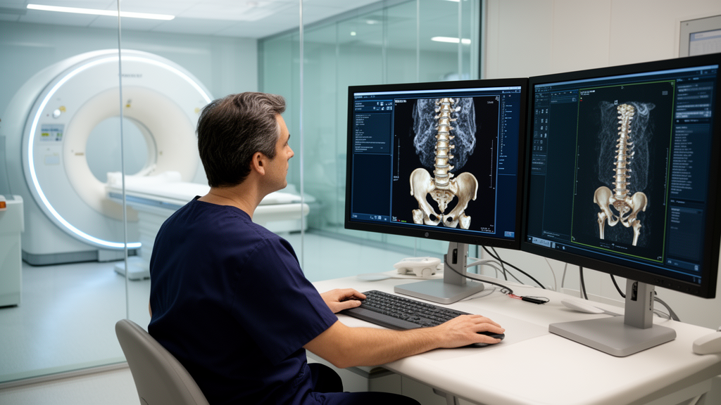
In orthopedic and trauma surgery, planning complex three-dimensional bone deformity corrections with external hexapod frames requires accurate anatomical measurements while exposing patients to minimal diagnostic radiation. A retrospective clinical study conducted at the BG Trauma Center Tübingen has demonstrated that an ultra-low-dose computed tomography protocol achieves a thirtyfold reduction in radiation dose compared to standard computed tomography, while maintaining full diagnostic adequacy for preoperative planning. Published in the Archives of Orthopaedic and Trauma Surgery, the investigation evaluated eighteen consecutive patients who underwent surgical correction of post-traumatic lower-extremity deformities using the Taylor Spatial Frame. The research team compared the effective radiation doses of conventional two-plane radiography, standard computed tomography, and an optimized ultra-low-dose protocol. While conventional radiographs delivered the lowest absolute exposure, ultra-low-dose computed tomography provided sufficient image quality to define crucial mounting parameters and anatomical axes at an average effective dose of only 4.61 microsieverts.
Modern hexapod fixators represent exceptionally precise surgical instruments for limb lengthening and complex multiplanar deformity corrections. These devices rely on computer-assisted planning software, such as the web-based SMART-TSF application, to calculate strut adjustments that gradually realign malaligned bone segments over several months. To generate an effective correction schedule, surgeons must determine the spatial relationship between the reference ring and the bone, identifying exact mounting parameters, deformity parameters, and the center of rotation and angulation. Traditionally, planning relies on orthogonal radiographs taken in anteroposterior and mediolateral projections. However, conventional radiographic projections remain vulnerable to perspective distortion, geometric divergence, and visual obscuration caused by the bulky metallic rings and struts of the hexapod frame. Such projectional inaccuracies frequently compromise measurement precision, leading to residual deformities and requiring repeated radiographic exposures that progressively increase the patient’s cumulative radiation burden.
Although standard computed tomography eliminates projection artifacts and provides superior three-dimensional visualization of osseous structures, its substantial radiation exposure has historically restricted routine application for deformity planning, especially among young patients. To evaluate whether low-dose scanning could reconcile diagnostic accuracy and radiation safety, researchers examined a consecutive cohort of twenty-eight patients treated with the Taylor Spatial Frame at the Level I trauma center between March 1, 2022, and August 31, 2024. The investigative protocol applied strict inclusion criteria, admitting only individuals for whom standardized conventional radiographs, standard computed tomography scans, and ultra-low-dose computed tomography scans of the lower extremity were available. Ten patients lacking one or more modalities were excluded, resulting in a final study group of eighteen patients comprising fifteen men and three women with a mean age of 48.5 years, spanning from 22.0 to 72.8 years.
All computed tomography examinations were performed on a 128-slice single-source scanner, the SOMATOM Definition Edge from Siemens Healthineers. The standard clinical protocol for lower-extremity computed tomography utilized established scanning parameters designed for detailed musculoskeletal imaging without iterative reconstruction algorithms. The scanner operated at a tube voltage of 120 kilovolts and a tube current-time product of 95 milliampere-seconds. The examination employed a table pitch of 0.8, a gantry rotation time of 1.0 second, and a detector collimation of 128 by 0.6 millimeters. Reconstructed axial slices were generated at a thickness of 2 millimeters using a very sharp B75h reconstruction kernel displayed in a dedicated bone window. In clinical practice, standard scans were performed when surgeons needed to evaluate specific clinical indications, such as monitoring the structural progress of mineral calcification within the distraction callus during ongoing limb realignment.
For the ultra-low-dose computed tomography examinations, investigators adapted an established clinical protocol previously validated for lower-extremity torsional assessments. Using the same 128-slice scanner, the team reduced the tube voltage to 80 kilovolts and shortened the gantry rotation time to 0.5 seconds with a pitch of 1.0. Detector collimation remained 128 by 0.6 millimeters. To minimize exposure dynamically, the team utilized automatic tube current modulation, CARE Dose4D, configured with a low reference setting of 20 milliampere-seconds tailored for bone structures. Furthermore, the protocol integrated raw-data-based iterative image reconstruction using the Sinogram Affirmed Iterative Reconstruction algorithm, designated SAFIRE, at strength level 3. Images were reconstructed at a slice thickness of 2 millimeters utilizing a medium-sharp I50f kernel, producing axial, coronal, and sagittal cross-sections as well as three-dimensional models displayed in a bone window.
Conventional radiographic examinations were completed using a digital X-ray unit, the Fluorospot Compact system from Siemens Healthineers, following the hospital standard protocol for the lower leg and knee. Radiographs were acquired with the patient supine on the examination table using an under-table grid and automatic exposure control. Both anterior-posterior and medial-lateral projections were captured using standardized reference settings of 60 kilovolts and 4 milliampere-seconds. To determine total radiation exposure, the research team summed the dose-area product of the anterior-posterior and lateral views. This combined dose-area product was multiplied by a validated region-specific conversion factor for knee radiography, in accordance with the 2011 British Health Protection Agency report and Publication 103 of the International Commission on Radiological Protection. This conversion incorporated tissue-specific weighting factors of anatomical structures within the examination field to accurately estimate effective dose in microsieverts.
Calculating effective radiation doses for standard and ultra-low-dose computed tomography involved a rigorous mathematical model accounting for patient geometry and scanner output. On axial reconstructions, examiners measured the maximum anatomical diameter of the lower leg along anterior-posterior and medial-lateral axes. Because both lower extremities are typically positioned within the scan field during lower-leg computed tomography, effective patient diameter was calculated as the square root of the product of these perpendicular measurements multiplied by two. The team matched this diameter to size-specific conversion factors outlined in American Association of Physicists in Medicine Report 204 using the implemented 32-centimeter phantom model. Multiplying the volume computed tomography dose index by this conversion factor yielded the Size-Specific Dose Estimate. Multiplying this estimate by scan length produced adjusted dose-length products, which were multiplied by the specific conversion factor for knee examinations to determine effective doses.
Statistical evaluation revealed profound, statistically significant differences in effective radiation dose across the three modalities. Inferential analysis using repeated-measures analysis of variance with Greenhouse-Geisser correction demonstrated an omnibus effect of F(1.002, 17.023) = 302.5 (p < 0.001) with a partial eta-squared of 0.95. Pairwise comparisons with Bonferroni correction showed that standard computed tomography imparted an average effective dose of 150.8 microsieverts, ranging from 94.85 to 224.6 microsieverts with a standard deviation of 35.81 microsieverts. In contrast, ultra-low-dose computed tomography produced a mean effective dose of 4.61 microsieverts, ranging from 2.97 to 6.68 microsieverts with a standard deviation of 1.00 microsievert, representing a significant 146.2 microsievert reduction and an approximate thirtyfold dose decrease (p < 0.001). Conventional radiography yielded the lowest mean effective dose at 1.41 microsieverts, with values between 0.66 and 2.53 microsieverts and a standard deviation of 0.60 microsieverts, differing significantly from ultra-low-dose computed tomography by 3.2 microsieverts.
Importantly, the dramatic dose reduction achieved by ultra-low-dose computed tomography did not compromise the diagnostic utility of the examinations. To assess clinical applicability, the researchers conducted a blinded, randomized evaluation of all ultra-low-dose scans. The review panel comprised two experienced specialists: a senior orthopedic surgeon with twenty-two years of experience in surgical deformity correction and a musculoskeletal radiologist with eleven years of experience. Both specialists rated datasets dichotomously as adequate or inadequate for assessing cortical bone alignment and determining reference frame mounting parameters. The reviewers judged one hundred percent of the ultra-low-dose computed tomography datasets to be diagnostically sufficient for surgical planning. The cross-sectional scans enabled reliable identification of essential bony landmarks, while multiplanar reconstructions in axial, coronal, and sagittal planes, alongside three-dimensional models, provided spatial orientation unattainable with conventional planar radiography.
Placing these measurements into clinical and environmental context highlights the safety profile of the low-dose protocol. In Germany and across Western Europe, the average individual receives an annual natural background radiation dose of approximately 2 millisieverts from cosmic, terrestrial, and radon sources, with medical applications adding another 2 millisieverts for a total baseline of around 4 millisieverts per year. The effective dose of 4.61 microsieverts from ultra-low-dose computed tomography represents a tiny fraction of this background exposure, comparable to just a few days of ordinary living. Although three times higher than the 1.41 microsieverts of two-plane radiography, ultra-low-dose scanning eliminates projection distortion and superimposition artifacts that often necessitate repeat X-rays. By delivering high diagnostic precision at a fraction of standard computed tomography exposure, the protocol complies with the ALARA principle—keeping exposure as low as reasonably achievable—while safeguarding patients undergoing long-term deformity treatment.
Despite these encouraging results, the authors highlighted several methodological limitations. The evaluation of image quality relied on subjective dichotomous ratings by two clinical specialists rather than objective physical measurements of image noise, contrast-to-noise ratios, or spatial resolution, which are currently being evaluated in ongoing investigations. In addition, the retrospective study design and modest sample size of eighteen patients from a single trauma center limit immediate generalizability across different institutions and scanner platforms. Most importantly, the study focused on imaging parameters and planning adequacy without directly measuring final clinical outcomes, such as achieved correction accuracy, complication rates, or subsequent revision procedures. The research team is systematically collecting prospective follow-up data to determine long-term clinical efficacy, while examining emerging radiological technologies including photon-counting computed tomography and artificial-intelligence-assisted reconstruction algorithms that may allow further radiation dose reductions in orthopedic planning.
Subject of Research: Radiation dose comparison of imaging modalities for hexapod deformity correction planning
Article Title: Radiation exposure for planning deformity corrections with hexapods: comparison of conventional X-ray, standard CT, and ultra-low-dose CT
Article References: Schüll, D., Baumgartner, H., Krüger, B., Thelen, J., & Keller, G. (2026). Radiation exposure for planning deformity corrections with hexapods: comparison of conventional X-ray, standard CT, and ultra-low-dose CT. Archives of Orthopaedic and Trauma Surgery, 146(1), Article 345. https://doi.org/10.1007/s00402-026-06479-x
Image Credits: AI Generated
DOI: 10.1007/s00402-026-06479-x
Keywords: ultra-low-dose CT, computed tomography, hexapod fixator, Taylor Spatial Frame, deformity correction, radiation exposure, effective radiation dose, orthopedic trauma surgery, conventional radiography, dose-area product, size-specific dose estimate, iterative reconstruction, ALARA principle, mounting parameters, bone deformity planning, musculoskeletal radiology
News Source: Ophelia Keating. (November 14, 2026). Ultra-Low-Dose CT Cuts Deformity Planning Radiation Thirtyfold While Preserving Precision. Scienmag.