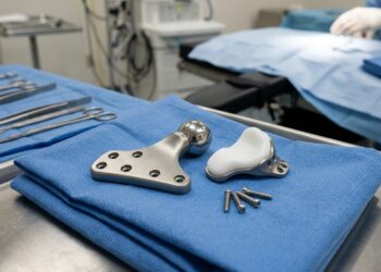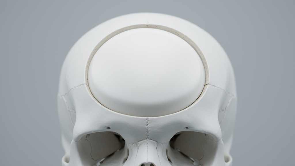
A retrospective review published in the Chinese Neurosurgical Journal found no statistically significant differences in postoperative complication rates between smooth and perforated polyetheretherketone (PEEK) implants used for cranioplasty. The study, conducted by researchers including Carmen A. Zavala and Mark J. Dannenbaum, analyzed data from 94 patients who underwent the procedure over a five-year period at a single institution. While the overall complication profiles were comparable, the analysis identified trends suggesting potentially higher rates of wound complications and infections in patients who received smooth implants. These findings, though not statistically significant, highlight the need for further investigation into how implant surface characteristics might influence clinical outcomes in neurosurgical reconstruction.
The goal of the procedure is to improve both functional and cosmetic outcomes for the patient. Historically, the gold standard for reconstruction has been the use of the patient’s own autologous bone flap. However, this method is associated with a high complication rate, which can reach up to 50% in some cases. Consequently, neurosurgeons have increasingly turned to synthetic materials to reduce these risks and improve the efficiency of the surgical process.
Among the synthetic options available, PEEK has become a popular choice due to its favorable physical properties. The material has tensile properties similar to bone, is thermodynamically stable, and demonstrates high resistance to radiation. Additionally, PEEK implants can be custom-designed using computer-aided design and manufacturing to fit specific patient defects, a process intended to decrease complications and enhance cosmetic results. Despite these advantages, questions remain regarding which specific type of PEEK implant offers the best post-surgical outcomes.
The study focused on two specific subtypes of PEEK implants: those with a smooth solid surface and those with perforated surfaces. The perforated implants featured 2 to 3 millimeter suture or drainage holes, which are typically used to tack up the dura or allow for drainage. In the current clinical practice, the choice between these two surface designs is often driven by surgeon preference rather than definitive evidence regarding their relative efficacy. The researchers aimed to determine if there were statistically meaningful differences in complication profiles between these two types, given that prior studies have suggested that surface structure can impact osseointegration and microbial susceptibility.
The research team queried electronic medical records from 2017 to 2022, initially identifying 529 procedures coded as cranioplasty. After applying exclusion criteria, which removed autologous cranioplasties, non-standard defects, and pediatric patients, 104 procedures remained. Ten of these were excluded due to incomplete data, leaving a final cohort of 94 patients. Of these, 45 received smooth implants and 49 received perforated implants. The demographic data showed that the two groups were comparable in age, comorbidities, and the primary indication for the initial craniectomy, with trauma being the most common reason for the initial surgery in both groups.
The primary results indicated that there were no statistically significant differences between the smooth and perforated implant groups regarding most measured outcomes. Rates of wound complications were 17.8% for smooth implants versus 8.2% for perforated implants, but this difference did not reach statistical significance. Similarly, postoperative infection rates were 17.8% for the smooth group and 8.2% for the perforated group, also without statistical significance. Other outcomes, including return to surgery, ventriculoperitoneal shunt placement, and implant removal, were statistically comparable between the two groups. Median hospital stay was three days for patients with smooth implants and two days for those with perforated implants, a difference that was not statistically significant.
Despite the lack of overall statistical significance, the multivariate logistic regression analysis revealed some notable trends. Smooth implants trended toward increased wound complications, postoperative infections, and ventriculoperitoneal shunt placement, although these associations did not meet the threshold for statistical significance. An interaction analysis did find a significant reduction in significant fluid collections for patients with smooth implants who had undergone craniotomies for trauma. This specific finding suggests that implant selection might be tailored to patient-specific clinical scenarios, such as the etiology of the initial craniectomy, to optimize outcomes.
The study also documented a rare and previously unreported case of malignant cerebral edema associated with a smooth PEEK implant. The patient was a 60-year-old male who had previously undergone resection of a meningioma and subsequent complications, including infection and hydrocephalus. After receiving a smooth PEEK cranioplasty, the patient experienced a seizure and rapid neurological deterioration, leading to brain death. Imaging confirmed diffuse malignant cerebral edema. While this is a devastating and rare complication, the authors noted that it has been previously reported with other implant materials, such as autologous bone and titanium, and that no association with implant type had been established prior to this case.
The authors emphasized that the chemically inert nature of PEEK can pose challenges for osseointegration, the process by which bone integrates with the implant surface. Previous studies have shown that smooth PEEK implants result in less osseointegration compared to those with porous surfaces or bioceramic coatings. The researchers suggested that this difference in integration might play a role in the observed complication profiles, although the impact of surface structure on clinical outcomes in cranioplasty remains sparsely described in the literature. The lack of statistically significant differences in this study supports the need for further clinical investigation into these mechanisms.
Limitations of the study include its retrospective design and the fact that it was conducted at a single institution. The sample size was relatively small, which may have limited the statistical power to detect smaller differences between the groups. Additionally, surgeon preference was a potential confounder, as a subset of attending surgeons preferred smooth implants while a larger group preferred perforated ones. The authors noted that a randomized clinical trial would be necessary to control for such factors and to provide more definitive evidence regarding the optimal choice of PEEK implant surface design for cranioplasty.
The findings contribute to the ongoing discussion about complication profiles in cranioplasty. Current literature suggests that factors such as patient age and the time interval between craniectomy and cranioplasty can influence outcomes. However, the optimal timing for the procedure remains debated, with some studies suggesting that earlier cranioplasty may increase the risk of hydrocephalus. In this study, there was no significant association between the length of time to cranioplasty and infection rate or wound revision. The results underscore the importance of continued research into the role of implant characteristics in reducing complication rates and improving patient safety in this common neurosurgical procedure.
As cranioplasty remains one of the most frequent neurosurgical procedures, the potential to reduce complication rates through optimized implant selection is a significant area of interest. The study’s identification of trends toward higher complications with smooth implants, along with the novel documentation of malignant cerebral edema, highlights the complexity of postoperative outcomes. While the current evidence does not support a definitive preference for one surface type over the other, the preliminary observations warrant further prospective studies to clarify these relationships and guide clinical decision-making in the future.
Beyond surface texture, the study detailed exact implant specifications: the CNC machine-milled medical-grade PEEK implants featured a 3-millimeter body with a 4-millimeter fixation area. In perforated models, 2-millimeter profusion holes were spaced 10 millimeters apart center-to-center, placed at least 15 millimeters from the edge. Although such perforations can anchor dural tenting sutures, the authors noted prior literature demonstrating that dural tenting does not significantly alter surgical outcomes, allowing the investigation to focus directly on surface architecture. Statistical calculations conducted in Python 3.9.18 and R 4.4.3 utilized Firth’s logistic regression to account for small sample sizes alongside Mann-Whitney U and Fisher’s exact tests.
The clinical assessment tracked four specific comorbidities: hypertension, diabetes, coronary artery disease, and coagulopathy. The authors also noted that no patients were on anticoagulation therapy at the time of surgery. Across the entire cohort, 11 patients (12%) developed wound complications or infections, culminating in eight implant removals, consisting of five smooth and three perforated implants. Microbiological cultures identified Staphylococcus aureus as the predominant pathogen across seven cases, alongside a single case of Propionibacterium acnes. Seven of these infected patients had originally undergone craniectomy for trauma and one for ischemic stroke. Multivariate modeling further demonstrated that craniectomies performed for intracranial hemorrhage were significantly associated with subsequent ventriculoperitoneal shunt placement (p = 0.021), whereas advancing patient age significantly predicted prolonged hospital stays of five or more days (p = 0.050).
Diagnostically, significant fluid collections were verified on postoperative computed tomography by neurosurgeons and radiologists, defined by the need for repeat monitoring scans or reoperation; these occurred in 2.2% of smooth implants compared to 10.2% of perforated implants. Regarding the rare case of malignant cerebral edema, the authors contextualized that this devastating condition is thought to stem from cerebral blood flow dysregulation or negative pressure drainage, factors historically linked to trephination syndrome and early surgical timing. The researchers also acknowledged unmeasured operative variables that could not be evaluated, including duraplasty methods, operative duration, and exact defect dimensions.
Subject of Research: Comparison of postoperative complications between smooth and perforated PEEK implants in cranioplasty
Article Title: Impact of PEEK implant surface design on postoperative complications in cranioplasty: a retrospective review
Article References: Zavala, C. A., Zima, L., Srinivasan, S., Neerukonda, S. V., & Dannenbaum, M. J. (2025). Impact of PEEK implant surface design on postoperative complications in cranioplasty: a retrospective review. Chinese Neurosurgical Journal, 11(1), Article 31. https://doi.org/10.1186/s41016-025-00417-3
Image Credits: AI Generated
DOI: 10.1186/s41016-025-00417-3
Keywords: PEEK implants, cranioplasty, neurosurgery, retrospective review, postoperative complications, implant surface design, wound infection, malignant cerebral edema, polyetheretherketone, craniectomy, osseointegration, clinical outcomes, skull reconstruction, medical devices, statistical analysis, ventriculoperitoneal shunt, trauma patients, single institution study, surgical revision, neurological outcomes
News Source: Ophelia Keating. (November 13, 2026). Study Finds No Major Difference in PEEK Cranioplasty Complications. Scienmag.