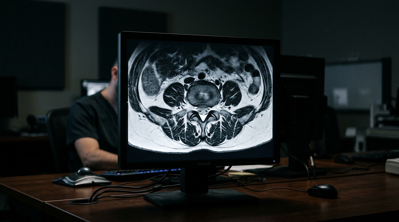
A systematic review analyzing 280 magnetic resonance imaging sequence applications across 274 published studies has revealed that three-quarters of investigations quantifying fatty infiltration in lumbar paraspinal muscles relied on conventional T1-weighted or T2-weighted imaging, despite substantial methodological variation and an absence of formal validation. The review, authored by Carla M. Zihlmann and colleagues and published in BMC Musculoskeletal Disorders, identified significant methodological heterogeneity across the literature evaluating muscle fat in the lumbar spine.
Fatty infiltration of the lumbar paraspinal muscles has been associated with spine degeneration and is extensively examined in relation to spinal disorders. While magnetic resonance imaging provides a non-invasive and clinically practical approach for evaluating muscle fat content, researchers noted that a comprehensive overview of existing quantitative imaging protocols, muscle segmentation techniques, and fat measurement approaches has been missing from the scientific literature. In addition, the review aimed to evaluate how machine-learning methods are currently being deployed within this research domain.
To evaluate the existing literature, the investigators carried out a systematic literature search across PubMed, Web of Science, and Scopus in October 2025. Following the screening of all retrieved publications against predefined inclusion and exclusion criteria, the authors systematically extracted data detailing the specific imaging sequences employed, the fat quantification methods utilized, reported reliability metrics, and participant cohort characteristics.
Among the 280 sequence applications evaluated across the 274 included publications, 209 applications, representing 75 percent, utilized either T1-weighted imaging or T2-weighted imaging. The remaining sequence applications incorporated chemical-shift magnetic resonance imaging techniques. The review noted that studies relying on T1-weighted and T2-weighted images predominantly investigated patient populations presenting with degenerative lumbar pathologies, whereas cohorts evaluated with chemical-shift imaging included both healthy individuals and participants diagnosed with spinal disorders.
Across the examined literature, manual segmentation remained the predominant approach researchers used to outline the lumbar paraspinal muscles of interest. Only a small subset of studies adopted machine-learning frameworks for muscle segmentation and subsequent fat quantification. Among the automated machine-learning approaches identified, models based on the U-Net architecture were the most frequently implemented by study investigators.
For studies implementing T1-weighted and T2-weighted images, investigators utilized multiple distinct approaches to quantify fatty infiltration within segmented muscle regions, including varied thresholding approaches, signal-intensity measures, and the direct manual outlining of adipose tissue. The reported reliabilities for these T1-weighted and T2-weighted fat quantification strategies varied substantially across the evaluated literature and were inconsistently documented. By comparison, chemical-shift techniques most frequently utilized the 6-point Dixon method, followed by 2-point and 3-point Dixon configurations.
The systematic review concluded that while T2-weighted imaging represents the most commonly used modality for evaluating paraspinal muscle fat, the applied quantification protocols vary considerably and lack rigorous validation, underscoring an urgent need for standardized and validated T2-weighted protocols to improve clinical utility and cross-study comparability. While chemical-shift techniques offer an inherently quantitative measurement based on resonance frequency differences between water and fat proton signals, they remain less commonly utilized in clinical research. The authors concluded that future research must prioritize establishing reliable automated segmentation methods to alleviate the substantial burden of manual muscle outlining.
The collaborative investigation was conducted by an author team comprising Carla M. Zihlmann, Ada Ayechu Abendaño, Sudeesh Arputham Dass, Mirko Blättler, McKinley Van Klei, Christoph E. Albers, Moritz C. Deml, Johannes T. Heverhagen, Ralph T. Schär, Keivan Daneshvar, and Ameet Aiyangar.
Chemical-shift techniques offer a quantitative evaluation based specifically on the difference in resonance frequencies between water and fat proton signals.
Subject of Research: Quantitative MRI techniques for measuring lumbar paraspinal muscle fatty infiltration
Article Title: Quantitative MRI techniques for measuring fatty infiltration in the lumbar paraspinal muscles: a systematic review
Article References: Zihlmann, C. M., Abendaño, A. A., Dass, S. A., Blättler, M., Van Klei, M., Albers, C. E., Deml, M. C., Heverhagen, J. T., Schär, R. T., Daneshvar, K., & Aiyangar, A. (2026). Quantitative MRI techniques for measuring fatty infiltration in the lumbar paraspinal muscles: a systematic review. BMC Musculoskeletal Disorders. https://doi.org/10.1186/s12891-026-10714-2
Image Credits: AI Generated
DOI: 10.1186/s12891-026-10714-2
Keywords: quantitative MRI, lumbar paraspinal muscles, fatty infiltration, systematic review, T1-weighted imaging, T2-weighted imaging, chemical-shift MRI, muscle segmentation, fat quantification, spine degeneration, spinal disorders, U-Net architecture, machine learning segmentation, 6-point Dixon method, 2-point Dixon method, 3-point Dixon method, signal-intensity measures, thresholding approaches
News Source: Ophelia Keating. (November 13, 2026). Systematic Review Finds Lumbar Paraspinal Muscle MRI Fat Quantification Methods Lack Validation. Scienmag.