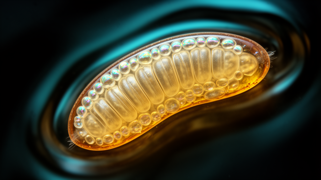
Researchers investigating the embryonic development of the German cockroach, Blattella germanica, have produced a detailed spatial and temporal timeline of early blastoderm patterning, cellularization, and segment formation. Unlike widely studied holometabolous insect models such as Drosophila melanogaster and Tribolium castaneum, this hemimetabolous species develops without forming a uniform blastoderm across the entire egg surface. The investigation, conducted using fluorescence microscopy, chromogenic in situ hybridization, hybridization chain reaction, and quantitative polymerase chain reaction, establishes that early cleavage nuclei condense directly onto localized lateral regions before fusing into a bipartite germband. In addition to mapping this direct-differentiating blastoderm, the authors found that key axis-patterning transcripts, specifically hunchback and caudal, are absent during initial cleavage stages, indicating that early polarity does not rely on maternally deposited messenger RNA gradients. Furthermore, the analysis demonstrates that segmentation in B. germanica occurs through two distinct regulatory dynamics, combining an almost simultaneous specification of gnathal and thoracic segments with a sequential, cycling addition of abdominal units from a posterior growth zone.
The German cockroach occupies an underrepresented phylogenetic position within the superorder Dictyoptera, belonging to the broader insect group Polyneoptera, which exhibits morphological and developmental features distinct from standard laboratory models. In typical model insects, early cleavage divisions produce a uniform blastoderm wherein energids evenly distribute across the egg periplasm before undergoing cellularization. In contrast, historical embryological literature described direct-differentiating blastoderms in certain hemimetabolous lineages, where dividing energids cluster in specific surface regions rather than dispersing homogeneously. Despite the organism’s widespread cosmopolitan distribution, gregarious habits, and established status as a pest and model for post-embryonic endocrinology, its earliest developmental phases had lacked a modern molecular characterization since light microscopy surveys from the 1970s. The authors sought to determine whether the absence of a uniform blastoderm alters the deployment of conserved developmental cascades, focusing on how cellular movements coordinate with the activation of transcription factors and segment polarity genes during the first fourteen percent of embryogenesis, when the primary body plan becomes morphologically established.
To observe early developmental progression under controlled conditions, the research team maintained an insect colony at twenty-five degrees Celsius and approximately forty percent relative humidity with a fourteen-to-ten hour dark-to-light cycle. Under these environmental parameters, total embryonic development lasts roughly twenty-five days, with initial germband formation spanning approximately three and a half days, representing fourteen percent of the total developmental trajectory. Female cockroaches produce eggs within protective cases called oothecae, carrying each case externally for three to four weeks until hatching. The deposition of eggs into an ootheca takes approximately twelve hours, creating a slight age gradient among individual embryos within a single egg case. To establish an accurate developmental baseline, researchers monitored laying females and designated the moment of complete egg case extrusion prior to its subsequent ninety-degree lateral rotation as time point zero. Staged embryos were subsequently dissected from removed oothecae using specific heating and cooling steps, followed by fixation in formaldehyde or paraformaldehyde depending on downstream staining requirements.
Using Sytox and DAPI nuclear staining, the investigators recorded nuclear division numbers and distributions across early germband formation, identifying two pronounced pulses of mitotic proliferation occurring between twenty-two to thirty hours and forty-four to fifty-three hours. Statistical comparisons of nuclear counts across seven- to eight-hour intervals confirmed significant increases in cell density across these developmental windows. Combining temporal counts with spatial observations, the authors partitioned early embryogenesis into four distinct morphological stages. Stage one, spanning days zero to one and zero to four percent of development, encompasses initial cleavage and blastoderm emergence. Following pronuclear fusion near the egg center, energids proliferate and migrate toward the egg periphery, appearing first along the ventral surface before reaching the dorsal margin. Immunofluorescence staining against phosphorylated histone H3 demonstrated dividing energids across the entire egg surface during stages one and two, showing that the non-uniform nuclear arrangement results from directed energid migration toward the ventral egg surface rather than localized, asymmetric cell divisions.
Stage two of development, occurring between days one and two and a half (four to ten percent of development), represents the syncytial blastoderm phase. During this stage, the dorsal hemisphere maintains a low density of energids, whereas the ventral hemisphere organizes into bilaterally paired regions of noticeably higher nuclear density. To identify the exact timing of membrane formation, the authors utilized wheat germ agglutinin staining paired with confocal fluorescence imaging. Examination of embryos collected twenty-four hours after ootheca extrusion revealed visible cell membranes in only one of five specimens, where agglutinin staining condensed closely around individual nuclei, whereas another embryo from the same egg case exhibited more nuclei but lacked detectable membrane envelopes. Embryos sampled between twenty-four and forty hours post-extrusion displayed pronounced membrane signals encircling nuclei, demonstrating that cellularization occurs midway through stage two, corresponding to four to six percent of total development, shortly after the first mitotic pulse concludes.
During stage three, extending from days two and a half to three and a half (ten to fourteen percent of development), the embryo transitions into the germ anlage or rudiment phase. In this window, the dorsal region resolves into two distinct nuclear populations: a group of larger, polyploid nuclei destined to form the extra-embryonic serosa, and a separate population of smaller cells resembling those within the germ rudiment. Concurrently, mitotic activity diminishes along the dorsal egg surface while ventral cell divisions persist, driving cellular condensation at the anterior edge where head lobes emerge. Stage four begins at three and a half days as accelerated cellular condensation causes the paired lateral plates to fuse along the ventral midline. Fusion initiates at the posterior extremity and advances anteriorly, culminating in the complete formation of a unified bipartite germband. This morphological sequence demonstrates how lateral plate migration establishes the preliminary body axis without relying on the extensive blastokinetic inversions observed in certain other hemimetabolous insects.
To evaluate embryonic polarization along the anterior-posterior axis, the team analyzed the transcription factor hunchback, a conserved anterior determinant, and the homeobox gene caudal, a conserved posterior organizer. In contrast to holometabolous dipterans where maternal mRNAs provide immediate positional cues, neither transcript was detected in early blastoderm embryos by chromogenic in situ hybridization or hybridization chain reaction. Quantitative polymerase chain reaction using primers validated against actin controls corroborated this absence, indicating that neither hunchback nor caudal is maternally deposited in B. germanica. Zygotic transcription of hunchback emerged two days post-extrusion (eight percent development), reaching a peak at three days (twelve percent development) before localizing along paired midline cells and head lobes. Zygotic caudal transcription commenced at day three and remained steady through day four, localizing to the posterior germband. These findings indicate that early axial polarization in hemimetabolous insects may operate independently of maternal transcription factor gradients, relying instead on structural egg cues.
The initiation of embryonic segmentation becomes detectable during stage three, as revealed by the complementary expression profiles of hedgehog, an arthropod segment polarity marker, and even-skipped, an upstream segmentation gene. Hybridization chain reaction and chromogenic probes demonstrated that even-skipped expression precedes hedgehog, initially manifesting as five simultaneous transverse stripes in the central germband between the head lobes and presumptive posterior addition tissue. These primary stripes define the territory spanning from the mandibular segment through the second thoracic segment. Shortly after head lobe condensation, hedgehog stripes appear individually in an anterior-to-posterior sequence. Despite this sequential appearance at the single-stripe level, the rapid formation of antennal, mandibular, maxillary, labial, and the first two thoracic segments occurs with minimal morphological change in the underlying germband. An eighth hedgehog stripe forms in the third thoracic segment just prior to head lobe fusion, after which the anterior even-skipped stripes progressively fade, illustrating an intermediate mode of anterior determination.
Posterior body organization shifts into a classical sequential segmentation dynamic as abdominal units emerge from a posterior segment addition zone. During abdominal elongation, individual segments sequentially initiate even-skipped expression before transitioning to hedgehog expression, with even-skipped signal gradually diminishing as hedgehog stripes intensify. This cycling turnover maintains approximately three active even-skipped stripes in the anterior portion of the segment addition zone throughout posterior extension. Alongside segmentation markers, the researchers tracked the basic helix-loop-helix transcription factor twist to assess mesoderm development. In stage three germ rudiments, twist expression appeared as punctate ventral-posterior foci, progressing during stage four fusion to occupy the lateral head lobes and posterior germband before covering most of the embryo. As segmentation advanced, twist expression became restricted to nascent limb primordia and newly formed segmental mesoderm, showing that despite lateral plate morphogenesis, early mesodermal marker distribution closely parallels patterns documented in Tribolium castaneum and Gryllus bimaculatus.
The findings illustrate that despite substantial divergence in early blastoderm architecture, core embryonic gene networks remain largely conserved between direct-differentiating polyneopterans and uniform-blastoderm model insects. The authors noted that the direct-differentiating blastoderm, characterized by regionalized nuclear emergence and localized surface retention, likely represents an evolutionary apomorphy of the superorder Polyneoptera. Furthermore, the transitional segmentation behavior observed in the third thoracic segment provides comparative evidence that tagmatization boundaries between thorax and abdomen reflect deeply conserved developmental transitions across insects. The researchers acknowledged methodological limitations, noting that live-cell imaging and fluorescent nuclear tracking could not be performed in this system, leaving migratory energid trajectories to be inferred from fixed-tissue assays. Additionally, while twist outlines early mesodermal territories, higher-resolution profiling will be needed to reconstruct the exact cell movements of ventral invagination. Nonetheless, this developmental atlas provides a quantitative framework for future functional studies employing modern genome-editing tools in Blattella germanica.
Subject of Research: Early embryonic development and gene expression dynamics in the German cockroach Blattella germanica
Article Title: Early embryonic development of the German cockroach Blattella germanica
Article References: Bar-Lev Viterbo, A., Wexler, J. R., Mayost Lev-Ari, O., & Chipman, A. D. (2024). Early embryonic development of the German cockroach Blattella germanica. EvoDevo, 15(1), Article 14. https://doi.org/10.1186/s13227-024-00234-2
Image Credits: AI Generated
DOI: 10.1186/s13227-024-00234-2
Keywords: Blattella germanica, German cockroach, early embryogenesis, blastoderm formation, direct-differentiating blastoderm, germband formation, cellularization, segmentation, hunchback, caudal, even-skipped, hedgehog, twist, Polyneoptera, Dictyoptera, tagmatization, segment addition zone
News Source: Drew Townsend. (November 13, 2026). German Cockroach Embryos Develop Distinct Blastoderms and Dual Segmentation Dynamics. Scienmag.