Diffuse axonal injury represents one of the most severe and elusive consequences of traumatic brain injury, frequently precipitated by high-velocity impacts, rapid angular acceleration, abrupt deceleration, and violent rotational forces. In fatal head trauma, such as that sustained during motor vehicle collisions or falls from height, structural damage inflicted upon the neural architecture often completely evades detection during conventional gross post-mortem examinations. This diagnostic invisibility has led forensic pathologists to designate diffuse axonal injury as a classic hidden lesion, where an anatomically unremarkable brain surface conceals catastrophic microscopic devastation within white matter pathways. Writing in the Egyptian Journal of Forensic Sciences, researcher Manal El Faham synthesized neuropathological, radiologic, and biomechanical evidence to demonstrate that establishing diffuse axonal injury in traumatic brain death demands a multimodal diagnostic strategy. By integrating advanced neuroimaging, specialized immunohistochemistry, and computational biomechanical simulations, death investigators can overcome the severe diagnostic blind spots that routinely compromise standard autopsy procedures and acute clinical assessments.
The underlying biomechanical foundation of diffuse axonal injury stems from the physical behavior of heterogeneous intracranial tissues subjected to rotational and translational motion. When head trauma induces sudden rotational acceleration or deceleration, intracranial tissues move at disparate rates due to variations in density and material properties. Consequently, mechanical shear strains become heavily concentrated along structural interfaces, particularly at the junction between the cerebral cortex and subcortical white matter, as well as within the corpus callosum, internal capsule, brainstem, and cerebellar peduncles. To clarify these internal dynamic events, forensic researchers increasingly employ finite element modeling, a computational engineering technique that maps complex brain geometry and material properties onto simulated mechanical impact vectors. By translating macroscopic cranial kinematics into localized tissue-level shear strain patterns, finite element modeling provides an objective physical explanation for how rotational forces concentrate destructive stress within deep axonal pathways even when external craniofacial impact markings appear minimal or absent.
At the cellular level, axonal pathology evolves across two distinct chronological phases comprising primary axotomy and secondary axotomy. Primary axotomy denotes the immediate mechanical disruption of axonal membranes and cytoskeletal structures occurring at the instant of physical impact. However, complete primary mechanical severance represents a relatively uncommon outcome. More frequently, damaged axons undergo secondary axotomy, an intricate biochemical and metabolic cascade developing over hours to days following trauma. Initial physical stretching impairs membrane integrity, triggering a pathological influx of calcium ions alongside mitochondrial dysfunction and oxidative stress. This intracellular calcium surge activates destructive proteolytic enzymes, specifically calpains and caspases, which degrade neurofilament frameworks and arrest axoplasmic transport. As transported proteins and cellular components accumulate proximal to injury sites, injured fibers progressively form swollen varicosities and retraction bulbs. This progressive sequence demonstrates that diffuse axonal injury is an evolving metabolic condition rather than an instantaneous static injury.
To categorize the spatial distribution and severity of these lesions, forensic neuropathology relies upon the tripartite grading system established by Adams and colleagues. This classification directly correlates the anatomical depth of tissue disruption with rotational force intensity and clinical outcomes. Adams Grade I represents the mildest category, defined by microscopic axonal injury confined to the cerebral white matter, corpus callosum, and brainstem in the complete absence of macroscopic lesions. Adams Grade II involves focal macroscopic lesions located within the corpus callosum, most commonly involving the posterior body and splenium, in addition to widespread microscopic pathology. Adams Grade III represents the most severe form, delineated by focal macroscopic lesions situated in the dorsolateral rostral brainstem and superior cerebellar peduncles.
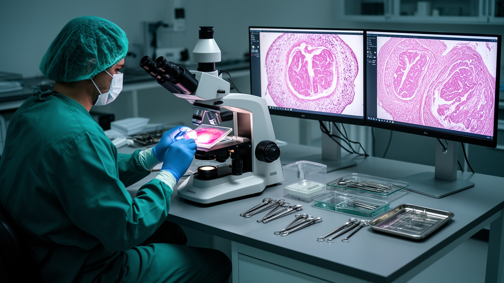
The post-mortem detection of diffuse axonal injury relies fundamentally upon targeted microscopic techniques, as routine staining frequently proves inadequate. White matter sections stained with conventional Hematoxylin and Eosin in cases with brief survival intervals often appear entirely unremarkable, obscuring early cytoskeletal injury and risking false-negative conclusions during standard gross autopsy. To expose these subtle disruptions, immunohistochemistry has become the diagnostic cornerstone in forensic pathology. Specifically, beta-amyloid precursor protein immunohistochemistry serves as the gold standard for histological confirmation. Additional markers, including ubiquitin and neurofilament proteins, provide complementary diagnostic sensitivity regarding the extent of cytoskeletal damage, while high-resolution silver staining and scanning electron microscopy retain diagnostic value for visualizing axonal retraction bulbs in more chronic investigations.
In both clinical trauma management and forensic post-mortem evaluations, radiological assessment reveals a substantial diagnostic discordance between imaging modalities. Computed tomography is universally deployed as the primary acute triage tool due to its rapid availability and effectiveness in detecting life-threatening intracranial hemorrhage and mass effect. However, computed tomography lacks sensitivity for non-hemorrhagic axonal shearing, frequently presenting normal findings or only non-specific cerebral edema. This limitation generates a critical clinicopathological discrepancy, where a seemingly normal scan is misinterpreted as an absence of injury despite severe underlying structural disruption. Conversely, magnetic resonance imaging provides far superior sensitivity, making it the preferred radiological modality. Conventional T2-weighted and fluid-attenuated inversion recovery sequences identify hyperintense lesions within the corpus callosum and brainstem, while gradient-recalled echo and susceptibility-weighted imaging sequences specifically detect microhemorrhages associated with shearing.
Advanced quantitative neuroimaging techniques have expanded the capacity to evaluate white matter microstructural integrity in traumatic brain injury. Diffusion tensor imaging quantifies white matter organization by measuring parameters such as fractional anisotropy. In addition, quantitative susceptibility mapping and diffusion kurtosis imaging provide heightened sensitivity for microhemorrhages and non-hemorrhagic lesions that elude standard scans. Nevertheless, practical limitations persist in medicolegal practice, as access to advanced modalities remains constrained across many jurisdictions. Furthermore, post-mortem magnetic resonance imaging faces distinct interpretive challenges, as post-mortem cooling and decomposition gas formation produce radiological artifacts that can mimic or obscure traumatic lesions. Consequently, radiological findings must always be interpreted in close conjunction with clinical context and targeted neuropathological evidence.
In pediatric forensic pathology, diffuse axonal injury represents an indispensable diagnostic marker, particularly in evaluations of suspected abusive head trauma in infants and young children. These biomechanical characteristics render infants exceptionally susceptible to rotational and acceleration-deceleration forces, permitting lower-magnitude trauma to generate significant axonal shearing. In suspected abuse cases, axonal damage frequently coexists with subdural hemorrhage, retinal hemorrhages, and cerebral edema. Identifying Grade II or Grade III diffuse axonal injury within the corpus callosum or brainstem provides crucial objective evidence of high-velocity rotational strain. This objective finding assists forensic pathologists in differentiating inflicted high-energy trauma from common household accidental falls from standing height, which typically produce focal contusions rather than deep axonal disruption. However, ongoing debate regarding shaking versus impact forces underscores the necessity of balanced multidisciplinary evaluations.
From a broader medicolegal perspective, establishing the presence and chronological evolution of diffuse axonal injury provides vital evidence for reconstructing death mechanisms and determining legal responsibility. Immunohistochemical detection of beta-amyloid precursor protein accumulation plays a central role in establishing injury timelines, enabling death investigators to determine a minimum post-injury survival interval to corroborate or refute witness accounts. However, forensic experts must approach pathological interpretation with substantial caution, as axonal retraction bulbs and swellings are not pathognomonic for trauma alone. Non-traumatic insults, including severe cerebral hypoxia, global ischemia, and metabolic encephalopathies, can induce identical disruptions in axoplasmic transport, producing histological mimics. Differentiating these mimics is particularly challenging in complex deaths involving both mechanical trauma and prolonged cardiopulmonary resuscitation. Expert testimony regarding diffuse axonal injury therefore requires careful communication of both diagnostic utility and intrinsic limitations, avoiding overinterpretation to prevent legal challenges and ensure the integrity of judicial outcomes.
To standardize forensic evaluations, El Faham synthesizes a multimodal diagnostic framework integrating biomechanical reconstruction, advanced neuroimaging, and beta-amyloid precursor protein histopathology evaluated through the Adams grading system. However, several inherent constraints must be recognized. A notable practical challenge is the temporal diagnostic gap, where instantaneous death prevents the metabolic accumulation of beta-amyloid precursor protein, yielding potentially false-negative microscopic results despite extensive mechanical trauma. Furthermore, finite element models rely on idealized material assumptions that represent approximations rather than exact biological reconstructions. To address these limitations, future directions emphasize correlating tissue pathology with peripheral blood biomarkers, such as neurofilament light chain and glial fibrillary acidic protein, which serve as objective indicators of axonal injury during early post-mortem intervals. Additionally, adopting ultra-high-field 7-Tesla magnetic resonance imaging, shifting toward imaging-guided targeted tissue sampling, and incorporating age-dependent brain geometry and material properties into personalized computational models will significantly advance diagnostic accuracy in traumatic brain death investigations.
Subject of Research: Multimodal forensic diagnosis and biomechanical evaluation of diffuse axonal injury in fatal head trauma
Article Title: The hidden lesion: multimodal forensic evaluation of diffuse axonal injury in traumatic brain death
Article References: El Faham, M. (2026). The hidden lesion: multimodal forensic evaluation of diffuse axonal injury in traumatic brain death. Egyptian Journal of Forensic Sciences, 16(1), Article 58. https://doi.org/10.1186/s41935-026-00556-3
Image Credits: AI Generated
DOI: 10.1186/s41935-026-00556-3
Keywords: diffuse axonal injury, forensic neuropathology, traumatic brain injury, beta-amyloid precursor protein, Adams grading system, abusive head trauma, rotational shear forces, secondary axotomy, susceptibility-weighted imaging, diffusion tensor imaging, finite element modeling, corpus callosum, retraction bulbs, fractional anisotropy, post-mortem magnetic resonance, neurofilament light chain, cerebral hypoxia mimics, medicolegal death investigation
News Source: Cassandra Pierce. (November 13, 2026). Multimodal Forensic Analysis Uncovers Hidden Diffuse Axonal Injury in Fatal Trauma. Scienmag.