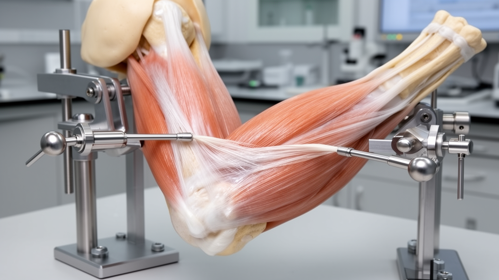
A laboratory investigation evaluating reconstructive strategies for posterolateral rotatory instability of the elbow has demonstrated that transferring a partial strip of the lateral triceps aponeurosis to the supinator crest provides measurable dynamic resistance against external rotational forces in lateral collateral ligament complex deficient joints. Conducting controlled biomechanical testing on nine fresh-frozen human donor elbow specimens, the research team found that this soft-tissue transfer significantly decreased pathologic external forearm rotation across multiple muscular loading configurations compared to an unconstrained, ligament-deficient state. The intervention altered the structural vector and functional lever arm of the triceps muscle, redirecting internal active forces to counteract displacing rotational torques. However, because the reconstruction did not completely restore native kinematic stability or match the rigid restraint achieved by formal transosseous anatomical repair, investigators concluded that the transfer functions primarily as a viable dynamic supplement or salvage option rather than a complete standalone replacement for primary ligamentous repair.
Posterolateral rotatory instability represents a disabling clinical pattern of elbow laxity that originates from functional disruption or insufficiency of the lateral collateral ligament complex, a specialized capsuloligamentous framework where the lateral ulnar collateral ligament serves as the primary static constraint against displacing external rotatory moments. In these demanding clinical revision settings, previous operative interventions often result in compromised lateral humeral condyle bone stock, multiple preexisting suture anchors, wide osseous defects, or crowded drill tunnels that physically preclude standard single-strand or double-strand anatomical graft anchoring. Consequently, researchers have sought alternative stabilization strategies that operate independently of compromised humeral attachment sites, prompting renewed scientific interest into how dynamic muscular contractions can actively reinforce joint articulation when static capsuloligamentous restraints are structurally deficient.
The biomechanical foundation of the procedure relies on anatomical geometry within the axial cross-section of the proximal radioulnar and humeroulnar articulations. Under normal physiological conditions, the lateral margin of the olecranon process serves as the native lateral insertion for the triceps apparatus, lying relatively close to the central axis of external forearm rotation. By contrast, the supinator crest on the proximal ulna is situated at a substantially greater radial distance from this instantaneous center of rotation. By mobilizing a one-centimeter-wide longitudinal strip of the lateral triceps aponeurosis while strictly preserving its proximal continuous attachment to the muscle belly and redirecting its distal terminal insertion to the isometric center of the supinator crest, the surgical configuration substantially elongates the effective moment arm. When muscular tension develops within the triceps, this enlarged mechanical lever arm generates an amplified internal restoring moment that directly opposes external rotatory displacement of the ulna relative to the distal humerus.
To rigorously test this mechanical hypothesis, the investigators designed a controlled in vitro testing apparatus using nine fresh-frozen upper extremity specimens obtained from seven female and two male human donors who had a mean age of 81.3 years, with a standard deviation of nine years and a range spanning from 68 to 99 years. After thawing for twenty-four hours at room temperature and undergoing comprehensive radiographic and manual physical screening to exclude joints exhibiting preexisting degenerative disease, traumatic injuries, surgical implants, or motion stiffness, each specimen was prepared by transsecting the proximal humerus to a length of fifteen centimeters. The proximal two centimeters of the humeral shaft were stripped of soft tissue and embedded securely in polymethylmethacrylate casting resin. Each prepared elbow was subsequently mounted at ninety to one hundred degrees of flexion and full forearm supination in a specialized testing rig secured within a servohydraulic materials testing machine.
Kinematic evaluation utilized a high-resolution three-dimensional optical tracking system recording positional coordinates at one hundred hertz from rigid intraosseous marker arrays rigidly fixed into both the humerus and the ulna. The experimental protocol imposed a standardized cyclic external rotation torque of 0.5 Newton meters directly to the proximal ulna via an olecranon hook passing through a bicortical tunnel at the articular level, pulling at a loading rate of seven Newtons per second for sixty consecutive cycles per condition. To prevent soft-tissue failure while maintaining reproducible joint displacement, an opposing counterweight system generating 0.3 Newton meters of internal rotation returned the forearm to its baseline starting position after each rotational deflection. Physiological muscle stabilization was simulated by routing freely suspended static calibration weights along the anatomical lines of action of the biceps and triceps brachii tendons across six predetermined combined loading levels, ranging from completely unloaded states up to four kilograms of triceps tension combined with half a kilogram of biceps countertension.
Because the progressive surgical preparation required irreversible anatomical alterations, each joint underwent sequential testing across four designated states in a non-randomized order: the native intact baseline articulation; an unstable state created by detaching the lateral collateral ligament complex and the common extensor origin from the lateral epicondyle; a repaired state following anatomical transosseous locking suture reconstruction; and finally the experimental tendon transfer state, wherein the repair sutures were completely released and the one-centimeter lateral aponeurotic triceps strip was shuttled through deep intermuscular planes and anchored to an ulnar drill tunnel with a cortical fixation button under manual tension. Statistical evaluation was conducted using a two-factor repeated-measures analysis of variance evaluating the unstable, repaired, and transferred conditions across all six muscular loading levels, while a separate one-factor repeated-measures analysis evaluated baseline performance against the native unloaded reference joint.
Quantitative analysis revealed a highly statistically significant main effect for the surgical intervention on external forearm rotation across the combined loading conditions, yielding an F-ratio of 33.31 with two and sixteen degrees of freedom, an associated p-value of less than 0.001, and a partial eta squared of 0.81. Across all tested muscle-loading states, the estimated marginal mean external rotation was 10.56 degrees with a standard error of 1.20 degrees in the unstable joint, which decreased significantly to 7.18 degrees with a standard error of 1.07 degrees following partial triceps aponeurosis transfer, representing a significant mean reduction of 3.38 degrees at a p-value of 0.005. Transosseous anatomical repair of the lateral collateral ligament complex demonstrated even tighter rotational constraint, producing a marginal mean rotation of 4.05 degrees with a standard error of 0.52 degrees, which represented a statistically significant 3.13 degree reduction compared to the tendon transfer at a p-value of 0.012.
Muscular loading independently exerted a pronounced stabilizing effect across all experimental conditions, with the repeated-measures model demonstrating a Greenhouse-Geisser corrected F-ratio of 77.00 with 1.26 and 10.11 degrees of freedom, a p-value of less than 0.001, and a partial eta squared of 0.91. When pooling across all surgical states, estimated marginal mean external forearm rotation progressively decreased from an initial high of 13.37 degrees with a standard error of 1.26 degrees under completely unloaded muscle conditions down to 2.42 degrees with a standard error of 0.40 degrees under the highest combined loading state of 0.5 kilograms of biceps tension and 4.0 kilograms of triceps tension. The interaction between surgical intervention and loading condition was also statistically significant, producing an F-ratio of 24.45 with 2.31 and 18.50 degrees of freedom, reflecting the fact that dynamic muscle tension exerted differential stabilizing magnitudes depending upon the underlying mechanical integrity of the lateral capsuloligamentous restraints.
At the completely unloaded baseline state involving zero kilograms of applied muscle tension, the separate one-factor repeated-measures analysis demonstrated significant variance among all four joint conditions, with an overall F-ratio of 37.96 and a p-value below 0.001. Intact native joints exhibited an external rotation of 4.81 degrees with a standard deviation of 1.80 degrees, which increased sharply to 19.95 degrees with a standard deviation of 5.02 degrees upon complete detachment of the lateral collateral ligament complex and extensor origin. Following transosseous repair, external rotation returned to 6.00 degrees with a standard deviation of 2.15 degrees, a value not statistically different from the native joint baseline at a p-value of 0.985. In comparison, the unloaded triceps transfer state exhibited 14.14 degrees of rotation with a standard deviation of 6.23 degrees, which was significantly more lax than both native and repaired states, while differing from the completely unstable condition by 5.81 degrees, a contrast that narrowly missed formal statistical significance after Bonferroni adjustment with a p-value of 0.058.
The study authors highlighted several methodological boundaries that govern the interpretation of these biomechanical findings. Because this investigation utilized an in vitro, time-zero cadaveric design, the experimental protocol could not account for dynamic biological variables such as progressive tendon-to-bone healing, vascularized soft-tissue remodeling, graft stretching, or active neuromuscular sensorimotor feedback loops that regulate dynamic joint co-contraction in living patients. Furthermore, testing was conducted at a fixed elbow flexion angle of ninety degrees and full supination under uniplanar rotational loading, leaving joint behavior across broader functional arcs of flexion and extension unmeasured. The advanced chronological age of the donor tissue and the necessary sequential testing sequence, which introduced potential soft-tissue relaxation over repeated testing cycles, also represent important constraints. Consequently, researchers emphasize that while partial triceps transfer successfully recruits dynamic muscle forces to restrain subluxation, it should not replace standard anatomical reconstructive techniques, serving instead as a targeted salvage option when compromised humeral bone precludes conventional static ligament repair.
Subject of Research: Biomechanical evaluation of partial triceps tendon transfer for dynamic stabilization in lateral collateral ligament deficient elbow models
Article Title: Partial triceps tendon transfer for dynamic stabilization in posterolateral rotatory instability of the elbow: a biomechanical study
Article References: Pucher, C.-J., Lanzerath, F., Kahmann, S. L., Hackl, M., Müller, L. P., & Leschinger, T. (2026). Partial triceps tendon transfer for dynamic stabilization in posterolateral rotatory instability of the elbow: a biomechanical study. Archives of Orthopaedic and Trauma Surgery, 146(1), Article 358. https://doi.org/10.1007/s00402-026-06520-z
Image Credits: AI Generated
DOI: 10.1007/s00402-026-06520-z
Keywords: elbow biomechanics, posterolateral rotatory instability, lateral collateral ligament complex, lateral ulnar collateral ligament, triceps aponeurosis transfer, dynamic stabilization, supinator crest, cadaveric study, orthopaedic surgery, joint kinematics, servohydraulic testing, rotational laxity, salvage procedure, optical motion tracking, repeated measures ANOVA, external rotation torque, Kocher interval
News Source: Ophelia Keating. (November 12, 2026). Partial Triceps Tendon Transfer Reduces Elbow Rotatory Instability in Biomechanical Study. Scienmag.