Published in EvoDevo, the study establishes a comprehensive 16-stage staging system for I. scapularis embryos and provides definitive evidence that early development proceeds through a syncytial phase, where nuclear division occurs without cell membrane formation, prior to the establishment of the blastoderm. This finding clarifies the morphological and temporal events that were previously missing from the scientific literature for this genus, which is the primary vector for Lyme disease in the eastern United States.
The study addresses a critical knowledge gap in tick biology, as previous research on early embryonic development in Ixodes species was largely absent or based on conflicting observations from other tick genera. While earlier studies on the cattle fever tick, Rhipicephalus microplus, suggested that cellularization might occur as early as 24 hours after egg laying, other investigations indicated a syncytial state lasting up to five days. The authors aimed to fill this gap to support the development of functional genomics tools, specifically embryo injection protocols required for creating stable germline transformants. Effective genetic transformation requires precise knowledge of when cell membranes form, as introduced genetic material must access the nucleus before cellularization to ensure integration into the primordial germ cells.
To characterize these early events, the team developed a staging system consisting of 16 distinct stages, labeled 0 through 15, based on unique morphological features observed via DAPI staining and time-lapse imaging. The embryos were maintained at 20 degrees Celsius, a standard condition for tick development. Stage 0, defined as less than 5 hours after egg laying, was a novel addition to the staging scheme, characterized by the absence of visible nuclei within the homogenous yolk. This stage was established to account for the earliest time points that were not characterized in previous studies of other tick species, providing a baseline for subsequent developmental observations.
Stage 1 began approximately 4 to 5 hours after egg laying, when a single nucleus became visible in the center of the egg via DAPI staining. Time-lapse imaging revealed that the first cleavage occurred deep within the yolk, with four cleavage energids, defined as nuclei and their surrounding cytoplasm, becoming visible between 6 and 12 hours after egg laying. The timelapse data also suggested that syngamy, the fusion of male and female pronuclei, likely occurred within the first hour after egg laying. The initial cleavages appeared to be synchronous, a pattern that contrasted with the asynchronous divisions observed in later stages.
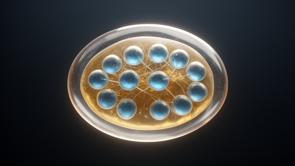
Stages 2 and 3, spanning from 12 hours to 3 days after egg laying, were characterized by rapid nuclear division without cellularization. During stage 2, the energids divided rapidly in the center of the egg and began migrating toward the periphery. By stage 3, the nuclei had reached the surface of the egg, and cleavage continued in a meroblastic manner, meaning nuclear division occurred without cytokinesis. The energids were connected to the cortical periplasm by thin projections along yolk columns, resulting in a multinucleated syncytial embryo. Unlike the syncytial blastoderm of Drosophila melanogaster, where nuclei are part of a continuous cytoplasmic layer, the tick energids were separated by densely packed yolk granules, and neighboring nuclei were not in mitotic synchrony.
Cellularization was confirmed to occur during stage 4, between 4 and 5 days after egg laying. The researchers employed two independent methods to verify the formation of cell membranes. First, they injected live embryos with a dextran-conjugated fluorescent dye, Dextran-Alexa Fluor 647. In embryos at 2 days after egg laying, the dye diffused completely throughout the embryo, indicating the absence of cell membranes. At 3 days, the dye was more visible at the periphery, suggesting the initiation of cytokinesis. In contrast, at 4 days, the dye remained localized at the injection site, confirming that complete cytokinesis had occurred and cell membranes were present.
Second, the team used wheat germ agglutinin (WGA) conjugated to fluorescein isothiocyanate (FITC) to label cell membranes in fixed embryos. WGA binds to glycoproteins containing N-acetylglucosamine and sialic acid residues, which are predominantly found on plasma membranes. WGA labeling was observed at 4 to 5 days after egg laying, corroborating the results from the dye injection experiments.
The study also investigated the formation of the cumulus, a cluster of mesenchymal cells that plays a crucial role in establishing the dorsal-ventral axis in chelicerates. At stage 5, 6 to 7 days after egg laying, a distinct germ disc formed in one hemisphere of the egg. The researchers detected bone morphogenetic protein (BMP) activity in a cluster of cells beneath the surface epithelial layer using antibody staining against phosphorylated Mothers against dpp (pMAD). This detection confirmed the presence of cumulus cells, which express decapentaplegic (dpp), a BMP homolog, to induce BMP activity in overlaying epithelial cells. By stage 7, the pMAD staining was spread around the ventral germband and was no longer localized to a specific cluster.
Subsequent stages, 6 through 15, described the progression from gastrulation to larval hatching. The ventral germ band formed along the longitudinal axis by stage 7. Embryo inversion occurred during stage 8, internalizing the yolk and splitting the ventral germband into two halves. Segmentation of the germ band became apparent at stage 9, 13 to 14 days after egg laying, with visible grooves defining the prosomal and opisthosomal regions. It was only at this stage that the anterior and posterior ends became clearly apparent.
Stages 10 through 12 involved the extension of the germ band, budding of appendages, and the beginning of germ band retraction. Four pairs of legs and a pair of pedipalps emerged ventrolaterally from the germ band. The post-abdomen, a structure that adds segments to the posterior growth zone, moved from the dorsal to the ventral side of the embryo. Dorsal closure was initiated by day 17 as cells from the prosoma and opisthosoma migrated dorsally. Stages 13 through 15, spanning from 19 to 40 days, were characterized by ventral closure, formation of the rectal sac, and synthesis of the chitinous cuticle. The embryo darkened as the cuticle was deposited, and the larva eventually hatched by breaking the vitelline membrane.
The findings of this study have significant implications for the development of genetic tools in tick research. The authors suggest that targeting embryos before cellularization, specifically before 3 days after egg laying, may be the optimal window for germline transformation strategies. Transformation during the pre-cellularization stage could result in uniform integration across lineages, including germ cells, which is essential for creating stable transgenic lines. The study also highlights the differences between tick embryogenesis and that of insects, such as Drosophila, where cellularization occurs much earlier and is associated with different axis determination mechanisms.
Limitations of the study include the inability to track cellularization in real-time using WGA-FITC injection in live eggs, which was successful in other arthropods but not in I. scapularis. The authors attribute this to the slower developmental pace of the tick, which may lead to the degradation of the fluorescent probe. Additionally, while the study provides a detailed staging system, the precise molecular mechanisms underlying the syncytial phase and the regulation of cellularization remain to be fully elucidated. The data presented here, however, provides a robust foundation for future studies aimed at understanding the molecular basis of tick development and improving genetic transformation techniques.
The research underscores the importance of detailed embryological studies in vector species for advancing public health strategies. By providing a standardized staging system and clarifying the timing of cellularization, the study facilitates the development of new genetic tools that could be used to study host-pathogen interactions and potentially develop novel control measures for tick-borne diseases. The work represents a significant step forward in the field of tick biology, offering insights that were previously unavailable for the Ixodes genus.
Subject of Research: Early embryonic development in the tick Ixodes scapularis suggests syncytial organization and cellularization before blastoderm formation
Article Title: Early embryonic development in the tick Ixodes scapularis suggests syncytial organization and cellularization before blastoderm formation
Article References: Hinne, I. A., Ciccotti, H. R., Wudarski, J., Pham, M. N., Sharma, A., McVicar, M. M., Faustino, B., Nuss, A. B., Sharma, P. P., & Gulia-Nuss, M. (2025). Early embryonic development in the tick Ixodes scapularis suggests syncytial organization and cellularization before blastoderm formation. EvoDevo, 16(1), Article 4. https://doi.org/10.1186/s13227-025-00240-y
Image Credits: AI Generated
DOI: 10.1186/s13227-025-00240-y
Keywords: Early, embryonic, development, tick, Ixodes, scapularis, suggests, syncytial, organization, cellularization, blastoderm, formation
News Source: Drew Townsend. (November 12, 2026). Tick Embryo Study Reveals Syncytial Stage Before Cellularization. Scienmag.