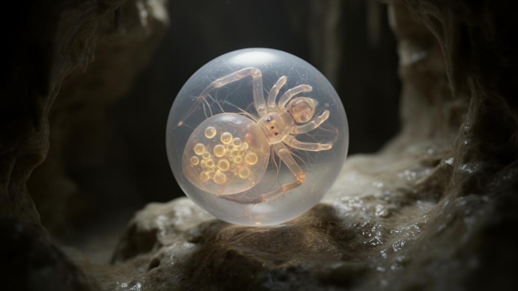
Developmental biologists investigating subterranean adaptation have documented the complete embryonic timeline and retinal gene expression dynamics of the troglophile cave spider Tegenaria pagana, establishing an embryological benchmark for understanding how sensory structures evolve and diminish in subterranean environments. Published in the journal EvoDevo, the investigation integrates confocal microscopy, fluorescent nuclear staining, and hybridization chain reaction protocols to track development from initial cleavage through hatching while mapping eight key genes within the retinal determination network. By resolving both the morphological stages of embryogenesis and the spatial expression patterns of visual regulatory factors in this eye-bearing spider, the research team constructed a foundational reference system for tracing the ontogenetic divergences that lead to blindness across cave-dwelling arachnids.
Subterranean caves represent ecologically distinctive environments defined by stable abiotic conditions, including the total absence of sunlight, constant temperatures, elevated humidity, and a lack of local primary photosynthetic production. Under these enduring aphotic constraints, colonizing fauna frequently evolve troglomorphic traits, prominently featuring the reduction or loss of eyes, cutaneous depigmentation, and elongation of appendages alongside compensatory enhancements in non-visual senses such as proprioception and chemoreception. While troglomorphic eye degeneration has been analyzed extensively in classic models such as the Mexican blind cavefish Astyanax mexicanus and the subterranean isopod Asellus aquaticus, comparative developmental data for arachnids have remained remarkably sparse despite their predatory roles in cave ecosystems. Furthermore, spiders exhibit diverse ocular architectures compared to insects, complicated by an ancestral whole-genome duplication event. Prior to this study, complete embryogenesis across more than fifty-two thousand described spider species had been documented from fertilization to hatching in only three taxa: Cupiennius salei, Parasteatoda tepidariorum, and Acanthoscurria geniculata, leaving cave spiders uncharacterized.
To establish the developmental trajectory of Tegenaria pagana under laboratory conditions, researchers collected twenty-seven adult females, nineteen males, and over twenty juveniles from Te’omim cave in Israel. Spiders were housed individually in containers measuring fifteen centimeters high and ten centimeters wide with autoclaved soil, stones, and moistened paper maintained at fifty percent humidity within dark incubators held between twenty and twenty-five degrees Celsius. Spiders received weekly rations of Drosophila melanogaster fruit flies. Controlled mating trials yielded one hundred forty-four successful copulations out of one hundred seventy-five pairings; pairs were kept in darkness for six to twelve hours after females constructed webs, with cannibalism occurring in rare instances where males were killed eight times and females twice. Gravid females typically deposited their first egg sac fourteen to twenty days following copulation. Egg sacs were collected within three days and incubated at twenty degrees Celsius to replicate cave conditions, allowing embryos to be monitored through time-lapse stereomicroscopy and preserved across development.
Embryonic morphology was characterized by dechorionating spherical eggs measuring five hundred to six hundred micrometers in diameter with bleach, followed by saline washes, formaldehyde-heptane fixation, methanol dehydration, and DAPI fluorescent staining for confocal imaging. The staging system, adapted from the fourteen-stage framework of the model Parasteatoda tepidariorum, begins with early intralecithal cleavages during Stage 1 from zero to ten hours after egg laying, wherein nuclear divisions proceed deep within homogeneous yolk before energids migrate toward the surface. During Stage 2, lasting eleven to thirty hours, energids populate the surface to establish a blastoderm of irregular polygonal cells, accompanied by deeper energids interpreted as putative vitellophages. Stage 3, occurring between thirty-one and forty-nine hours, exhibits blastopore formation as blastoderm cells shift asynchronously into a localized depression; notably, Tegenaria pagana lacks a distinct germ disc, resembling Cupiennius salei. During Stage 4, spanning fifty to seventy-two hours, blastopore cells ingress beneath the blastoderm to drive gastrulation, forming a surface protrusion that flattens into a primary thickening.
The middle phases of embryogenesis govern axial patterning and segment proliferation, beginning at Stage 5 between seventy-three and one hundred six hours as mesendodermal cells segregate into two discrete clusters. A larger cellular aggregate, the cumulus, elevates above the embryonic surface and migrates along an arc of one hundred to one hundred ten degrees across the egg, ultimately dissolving to designate the dorsal pole while the primary thickening marks the posterior pole. Stage 6, lasting one hundred seven to one hundred fourteen hours, initiates dorsal field formation through cell migration away from the primary thickening, which subsequently forms the caudal lobe. At Stage 7, extending from one hundred fifteen to one hundred eighteen hours, a dense C-shaped germ band forms across the ventral hemisphere, terminating posteriorly in a segment addition zone. During Stage 8, spanning one hundred nineteen to one hundred fifty hours, the germ band expands across three hundred degrees of the perimeter, resolving six prosomal segments alongside three sharply demarcated opisthosomal segments.
Subsequent organogenesis progresses through Stage 9, spanning one hundred fifty-one to one hundred eighty-five hours, as prosomal limb buds elongate outward while opisthosomal segments increase to five segments of uniform size, contrasting with Parasteatoda tepidariorum. At substage 9.2, internalized neural precursor cells appear as discrete point-like depressions within the precheliceral region, accompanied by the earliest morphological signs of the respiratory book lung system and a visible ventral sulcus. Stage 10, extending from one hundred eighty-six to two hundred twenty-three hours, brings neurogenic differentiation as the precheliceral region divides into paired lobes featuring kidney-shaped lateral furrows, while the stomodeum, paired labrum, pedipalp endites, and seven opisthosomal segments differentiate. Embryonic inversion unfolds during Stage 11 between two hundred twenty-four and two hundred thirty-five hours, as lateral opisthosomal growth internalizes the yolk and the segment addition zone extends into a tail-like post-opisthosoma. During this stage, chelicerae divide into bases and fangs, walking legs resolve into six segments, and an invaginating slit marks the developing book lungs.
The concluding morphological transitions encompass germ band retraction and structural enclosure, initiating at Stage 12 between two hundred thirty-six and two hundred sixty-one hours as the germ band retracts, driving the posterior abdomen away from the precheliceral lobes. Concurrently, walking legs adopt an interlocking arrangement with seven distinct segments, tergal plates internalize exposed yolk, and posterior opisthosomal segments form a recurved caudal tail. Stage 13, extending from two hundred sixty-two to two hundred ninety-seven hours, achieves dorsal closure as bilateral tergites fuse along the dorsal midline from posterior to anterior, generating the prosomal shield while three pairs of developing secondary eyes become detectable above the chelicerae alongside transient pedipalpal egg teeth.
To unravel the genetic regulatory mechanisms directing spider ocular development, the authors evaluated candidate Retinal Determination Network genes across embryonic stages 9.2 through 14 using hybridization chain reaction in situ hybridization paired with maximum likelihood phylogenetic verification. Although the classic master control gene Pax6 serves as the upstream initiator of retinal specification in Drosophila melanogaster, Pax6 orthologs could not be identified within the developmental transcriptome of Tegenaria pagana, mirroring prior findings across multiple spider families where Pax6 orthologs are absent or uncoupled from ocular tissue. Consequently, researchers focused expression profiling on eight conserved transcription factor homologs: Pax2.1, sine oculis 1, eyes absent, orthodenticle 2, dachshund 1, dachshund 2, Six3.2, and atonal 1.
The resulting gene expression profiles revealed both highly conserved core regulators and surprising spatial distributions that challenge assumptions regarding spider eye differentiation. The homeobox gene orthodenticle 2 was detected in both principal and secondary eye primordia by Stage 14, demonstrating that otd2 does not mediate eye-type divergence in Tegenaria pagana. Most notably, Pax2.1 was initially expressed within secondary eye fields before expanding symmetrically into all eye types by Stage 14, contradicting hypotheses that Pax2.1 serves as an exclusive functional replacement for Pax6.
Synthesizing these embryological and molecular observations, the study establishes that while gross morphogenetic milestones remain conserved across entelegyne spiders, significant variations govern developmental timing, ventral sulcus dynamics, opisthosomal segment proportions, and retinal gene deployment. The funnel-web genus Tegenaria encompasses one hundred thirty-five described species globally, including eleven species inhabiting subterranean cave systems across Israel that display an extraordinary evolutionary continuum ranging from fully eyed forms to species exhibiting progressive stages of ocular reduction and total blindness. By elucidating the normal developmental trajectory and retinal determination architecture of the troglophile Tegenaria pagana, this investigation furnishes an indispensable comparative baseline for future evolutionary developmental research. Identifying how regulatory networks falter, downregulate, or arrest during embryogenesis in closely related blind cave congeners will provide unprecedented mechanistic insight into the genetic changes driving troglomorphic sensory regression throughout subterranean evolution, shedding light on the broader evolutionary developmental pathways of arthropod adaptations.
Subject of Research: Embryonic development and retinal determination gene expression in the troglophile spider Tegenaria pagana
Article Title: Shedding light on the embryogenesis and eye development of the troglophile cave spider Tegenaria pagana C. L. Koch, 1840 (Araneae: Agelenidae)
Article References: Propistsova, E. A., Gainett, G., Chipman, A. D., Sharma, P. P., & Gavish-Regev, E. (2025). Shedding light on the embryogenesis and eye development of the troglophile cave spider Tegenaria pagana C. L. Koch, 1840 (Araneae: Agelenidae). EvoDevo, 16(1), Article 2. https://doi.org/10.1186/s13227-025-00238-6
Image Credits: AI Generated
DOI: 10.1186/s13227-025-00238-6
Keywords: Tegenaria pagana, embryogenesis, spider development, retinal determination network, troglophile, cave adaptation, eye reduction, sine oculis, eyes absent, dachshund genes, orthodenticle, Pax2.1, Entelegynae, Te'omim cave, gastrulation, blastoderm, chelicerate evolution
News Source: Juliet Wilcox. (November 12, 2026). Study Details Cave Spider Embryogenesis and Conserved Retinal Gene Networks. Scienmag.