Physiologists at Michigan State University have designed two complementary, low-cost physical models to help students grasp the competing physical forces behind the Reynolds number without relying on rote memorization or misinterpreting qualitative classroom observations. Published as a methods report in Frontiers in Physiology, the instructional framework developed by Victor Akinyemi, Heidi L. Lujan, and Stephen E. DiCarlo addresses a widespread and longstanding challenge in cardiovascular physiology education, where learners frequently memorize algebraic formulations while struggling to visualize how convective inertia and viscous damping interact in living circulation. Rather than attempting to serve as high-precision fluid-mechanics experiments or direct clinical simulators, the classroom apparatuses provide accessible visual analogies that demonstrate how physical flow variables are inherently coupled in dynamic circuits. The investigators deliberately structured these interactive learning activities around a predict-observe-explain sequence, establishing explicit learning targets centered on physical mechanism, conduit constraints, and the crucial scientific distinction between direct sensory observations and unmeasured diagnostic inferences.
In standard medical and physiological curricula, understanding why blood flow remains quiet and orderly under resting baseline conditions but turns noisy and disturbed during disease is essential for clinical practice. Learners typically encounter Poiseuille’s law, the continuity equation, Bernoulli’s relationship, and the dimensionless Reynolds number as isolated mathematical topics taught across separate lectures. Consequently, students often recall individual proportionality rules without comprehending how hydrostatic pressure, volumetric flow, linear velocity, conduit geometry, and dynamic viscosity interact within a single closed circuit. The classic Reynolds formulation expresses the ratio of convective inertial momentum to viscous damping forces in cylindrical conduit flow, incorporating fluid density, mean velocity, internal tube diameter, and dynamic viscosity. Because this dimensionless parameter cannot be observed directly, learners routinely struggle when analyzing complex pathologies such as severe anemia, polycythemia, heart valve stenosis, or arterial dilation, in which multiple hydrodynamic and biological variables shift simultaneously rather than altering one parameter at a time.
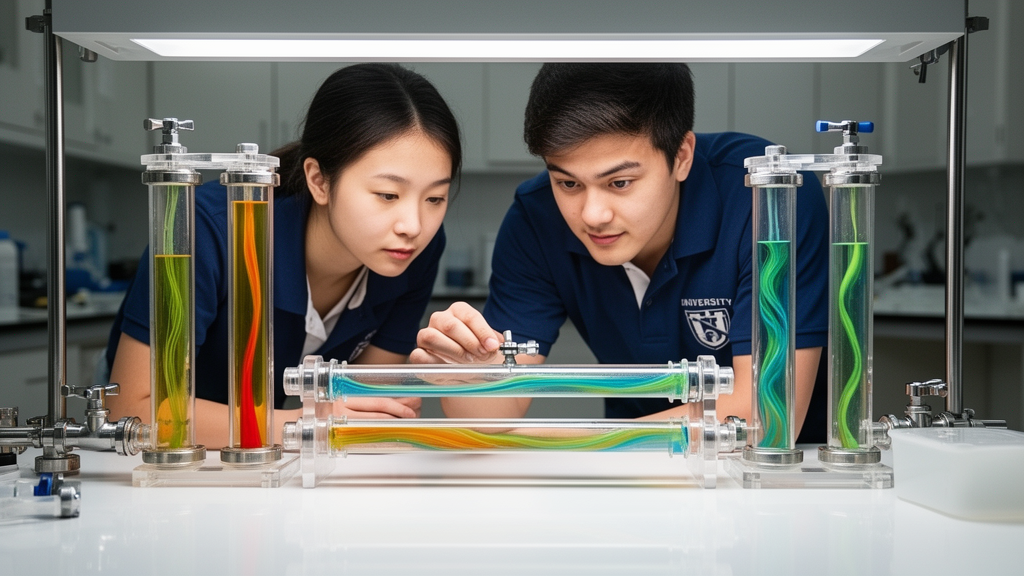
To render these abstract physical relationships visible without investing in expensive engineering equipment, the authors engineered their first demonstration using transparent fluid chambers and an adjustable dual-outlet aquarium air pump. In this bubble-chamber apparatus, flexible tubing equipped with inline non-return check valves routes air from the pump through submerged diffusers mounted within identical transparent cylinders repurposed from oxygen humidifier bottles. Each chamber lid features an air inlet, an atmospheric vent that remains open during operation to prevent progressive chamber pressurization, and an access port for adjusting test fluids. As air passes through the submerged diffusers, it generates an ascending, buoyant two-phase bubble plume. Students can directly observe and compare the vertical speed of rising bubbles, the transverse spreading width of the plume, lateral wandering, structural fluctuations, and the persistence of a central coherent column, using simple rulers or standard frame-by-frame video review to maintain consistent qualitative records across successive trials.
The bubble-chamber protocol deliberately imposes clear interpretive boundaries to guide classroom deductions. However, the authors stress that this buoyant two-phase gas-liquid mixture is governed by buoyancy, interfacial surface tension, bubble diameter, and coalescence, meaning its behavior cannot be equated with single-phase pipe flow or labeled as laminar or turbulent. In a second protocol, students evaluate fluid composition by observing plumes in a graded series of glycerol-water mixtures containing zero, twenty-five, fifty, and seventy-five percent glycerol by volume. Because adding glycerol simultaneously alters fluid viscosity, density, surface tension, and bubble dynamics, students are taught to treat the results as a fluid-composition comparison rather than attributing macroscopic plume changes solely to altered viscosity.
The complementary second demonstration tackles another frequent pedagogical hurdle by demonstrating that conduit diameter cannot be altered in a pump-driven system without altering downstream flow resistance, volumetric delivery, and exit velocity. This geometry-flow coupling apparatus utilizes a compact three-watt dual-outlet aquarium air pump connected to two independent, sealed water reservoirs containing commercial ultrasonic mist makers. A secondary gas port on each sealed reservoir conveys the generated water-droplet aerosol into horizontally aligned serological pipettes—one with a nominal five-milliliter capacity and the other with a nominal fifty-milliliter capacity—mounted at identical elevations and orientations. Commercial red light-emitting diode bicycle rear lights illuminate each aerosol discharge perpendicular to its direction of travel against a dark backdrop, creating high-contrast visual plumes without costly laboratory imaging systems. This arrangement allows students to examine the visible behavior of both discharge streams simultaneously under identical ambient conditions while maintaining stable tracer density and water levels.
Under the predict-observe-explain framework, students first predict how the two conduits will behave and then analyze the hydrodynamic principles underlying the observed difference. Because both lines are fed by an air pump rather than an idealized constant-flow source, modifying pipette geometry alters downstream flow resistance, which in turn alters the volumetric flow delivered through each branch. Although the continuity equation states that mean velocity equals volumetric flow divided by cross-sectional area, students discover that volumetric flow cannot be treated as a constant when pipe caliber changes. Furthermore, the visible aerosol plume represents an unconfined external free jet influenced by shear layers, entrainment, and room drafts, meaning a coherent plume cannot be interpreted as proof of laminar internal flow.
To reinforce rigorous scientific interpretation, the curriculum requires students to separate three distinct physical domains that are frequently conflated: internal conduit flow, the localized exit jet at the orifice, and the downstream free-spreading plume. An internal pipe Reynolds number is governed by internal tube diameter and mean internal velocity, whereas a jet Reynolds number depends on orifice diameter and exit velocity. The authors explain that calculating these actual numerical values would require calibrated branch-flow meters, direct fluid-velocity sensors, exact geometric profiling of the pipettes, and precise fluid density and dynamic viscosity values recorded at the ambient experimental temperature. Because commercial serological pipettes deviate from ideal cylindrical tubes and dual-outlet consumer pumps may exhibit internal asymmetry or branch interaction, the classroom model intentionally avoids reporting quantitative flow rates or Reynolds numbers.
In translating these physical demonstrations to cardiovascular physiology, the authors treat clinical conditions strictly as mechanistic analogies rather than exact biological replicas. For example, severe anemia reduces blood viscosity while compensatory mechanisms elevate cardiac output, both of which increase the relative importance of convective inertia and help explain benign systolic flow murmurs in individuals with structurally normal cardiac valves. Conversely, polycythemia elevates blood viscosity, which tends to reduce the Reynolds number for a given vessel geometry and velocity, even as it increases systemic vascular resistance and cardiac workload. In patients with arteriovenous fistulas, such as those surgically constructed for hemodialysis or arising pathologically in hereditary hemorrhagic telangiectasia and Paget disease of bone, blood bypasses high-resistance capillary networks. This low-resistance shunt reduces systemic vascular resistance, increases venous return, and elevates cardiac output, generating characteristic systolic murmurs and, when sufficiently severe, ultimately contributing to high-output heart failure.
Pathological vessel geometries similarly demonstrate why clinical hemodynamics cannot be understood by examining diameter or velocity in isolation. In valvular stenosis, a narrowed effective orifice generates a high-velocity jet with pronounced shear layers, flow separation, and downstream mixing that produce diagnostic murmurs, while regurgitant lesions produce high-velocity jets during distinct cardiac phases, including systole in mitral regurgitation and diastole in aortic regurgitation. Similarly, arterial aneurysms and post-stenotic dilations illustrate why localized lumen widening does not ensure smooth flow; although cross-sectional expansion decreases mean forward velocity, the abrupt geometric divergence provokes adverse pressure gradients, boundary-layer separation, recirculation vortices, and localized flow reversal. Importantly, instructors remind students that simple benchtop models omit vital cardiovascular characteristics, including cardiac pulsatility, elastic wall compliance, vascular branching, tortuous geometry, and the non-Newtonian, shear-dependent viscosity of whole blood, preventing any simplistic equivalence between rigid plastic tubes and living vascular beds.
To facilitate classroom adoption, the report outlines formative assessment strategies, including audience-response polling, think-pair-share discussions, and written prediction exercises aligned with five core learning objectives. Suggested debriefing questions challenge students to explain why nominal pump settings do not equal volumetric flow, why external jet coherence does not verify laminar internal conditions, and how simultaneous property shifts confound simplistic causal claims. The authors explicitly note that their publication describes qualitative instructional methods rather than a formal educational-outcomes investigation, as it includes no pre- and post-instruction testing cohorts, learning-gain metrics, or structured classroom survey datasets. Nevertheless, by pairing an aerated bubble column with an aerosol-illuminated dual-pipette circuit, the framework provides an accessible pedagogical platform that trains future clinicians and scientists to interpret physical analogies with discipline, respect model boundaries, and separate empirical observation from unsubstantiated inference.
Subject of Research: Qualitative physical classroom demonstrations for teaching Reynolds-number reasoning and variable coupling in cardiovascular physiology
Article Title: Making Reynolds-number reasoning visible: complementary classroom models for plume organization and geometry–flow coupling
Article References: Akinyemi, V., Lujan, H. L., & DiCarlo, S. E. (2026). Making Reynolds-number reasoning visible: complementary classroom models for plume organization and geometry–flow coupling. Frontiers in Physiology, 17, Article 2006049. https://doi.org/10.3389/fphys.2026.2006049
Image Credits: AI Generated
DOI: 10.3389/fphys.2026.2006049
Keywords: Reynolds number, cardiovascular physiology, fluid mechanics, classroom demonstrations, hemodynamics, active learning, variable coupling, laminar flow, Making, Reynolds-number, reasoning, visible
News Source: Drew Townsend. (November 12, 2026). Classroom Fluid Models Make Reynolds Number Reasoning Visible for Physiology Students. Scienmag.