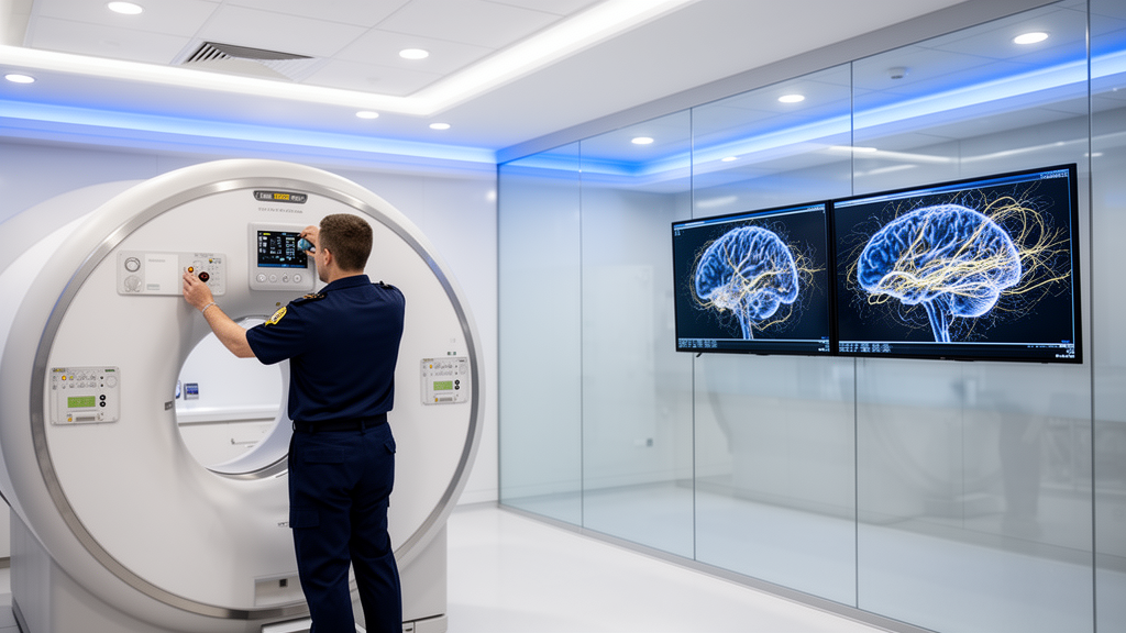
An editorial published in Frontiers in Neuroscience examines how methodological rigor across every stage of data collection, processing, and analysis is necessary to transform complex brain recordings into dependable clinical insights. Authored by Venkateswarlu Gonuguntla, Bhushan Borotikar, Kalyana C. Veluvolu, Michel Audette, Yubo Wang, and Zhiliang Wei, the article introduces a dedicated Research Topic titled Data-driven brain imaging and signal analysis: methods from acquisition to clinical insight within the journal’s Brain Imaging Methods section. The authors emphasize that rapid technological expansion across neuroimaging, electrophysiology, and advanced computation has substantially broadened scientific capabilities to examine the human brain. Modalities such as electroencephalography, intracranial electroencephalography, magnetic resonance imaging, diffusion imaging, and positron emission tomography provide intricate details regarding neural structure, function, dynamic activity, and molecular processes, while machine learning, deep learning, multimodal analysis, and distributed computing enable granular evaluations of these comprehensive datasets. However, the editorial cautions that deploying intricate algorithms does not inherently produce reliable results or meaningful clinical value. The ultimate integrity of data-driven neuroscience depends on the entire research pipeline, requiring accurate data acquisition, appropriate preprocessing, robust analytical strategies, and clinically relevant biological interpretation to ensure that computational findings translate effectively into practical scientific knowledge.
Reliable data acquisition constitutes the indispensable foundation for all computational modeling and analytical pipelines in neuroimaging. Empirical data collected through neural recordings and brain scans remain fundamentally sensitive to a broad spectrum of confounding influences, including specific participant characteristics, scan protocols, hardware configurations, operator habits, subject movement, acute physiological fluctuations, and clinical pathologies. When left unaddressed, these confounding variables impair both the reproducibility and the generalizability of downstream computational algorithms. To mitigate motion-induced distortions retrospectively, a study by Bricout and colleagues introduced an automated quality estimation framework using a three-dimensional convolutional neural network trained on synthetically motion-corrupted structural magnetic resonance imaging scans. By integrating this retrospective motion score into cortical morphometric evaluations, the researchers achieved measurable improvements in statistical model fit across multiple independent datasets. Their findings demonstrate that evaluating acquisition quality must not be treated as a secondary consideration, but rather as an essential, quantifiable parameter within quantitative neuroimaging workflows that directly impacts anatomical modeling accuracy.
Complementing retrospective motion adjustments, prospective standardization represents an equally critical requirement for clinical neuroimaging reproducibility. A study by Azeez and colleagues establishes that achieving dependable neuroimaging extends far beyond merely locking down standardized scanner parameters. Instead, true reproducibility relies on coordinated procedural workflows capable of accommodating the clinical needs of participants while simultaneously enforcing rigorous, uniform data collection and quality-control standards. By formally linking participant preparation routines, thorough staff training programs, and timely quality assessments, their proposed framework handles data fidelity as a collective responsibility embedded across the entire operational acquisition process. This prospective management prevents data quality from being treated as a remedial problem relegated exclusively to post-hoc preprocessing scripts. The editorial underscores that prospective operational workflows and retrospective motion correction algorithms serve complementary, reinforcing functions. Establishing explicit acquisition protocols, executing standardized quality audits, and maintaining exhaustive procedural documentation are mandatory prerequisites to ensure that collected neural datasets possess the fidelity needed to support meaningful downstream statistical computations.
A second foundational theme highlighted in the collection involves converting complex, high-dimensional neural measurements into representations that faithfully retain biologically and clinically relevant information. Because brain dynamics naturally unfold across widely distributed temporal and spatial scales, analytical success depends on selecting representations aligned with underlying neurophysiology. Investigating this dynamic landscape, Kuang and colleagues analyzed multi-frequency dynamic functional connectivity in schizophrenia using constrained tensor decomposition, demonstrating that functional connectivity patterns fluctuate systematically across both temporal intervals and distinct frequency bands. In a related investigation of temporal organization, Yan and colleagues evaluated frequency-specific electroencephalography microstates during ciprofol-induced transitions in consciousness, revealing that discrete microstate dynamics mirror changing conscious states. Furthermore, Wu and colleagues investigated stage-specific event-related potentials evoked during audiovisual facial emotion processing, identifying distinct electrophysiological responses that correlate with depressive tendencies. Collectively, these studies substantiate that mapping neural patterns across both temporal trajectories and spectral frequencies yields critical insights into the functional architecture of the brain and its direct connection to behavioral phenotypes.
Beyond group-level analyses, individual normative modeling provides an essential mechanism for characterizing patient-specific neurophysiological abnormalities in rare and heterogeneous clinical populations. Sharma and colleagues demonstrated this principle by constructing an age-referenced normative quantitative electroencephalography reference space, utilizing Mahalanobis distances to quantify individualized deviations in patients diagnosed with SNAREopathies. Their computational formulation successfully linked these individualized statistical deviations to distinct clinical severity dimensions, highlighting how normative reference spaces allow clinicians to contextualize rare neurodevelopmental disorders against healthy developmental trajectories. Such normative frameworks provide a principled baseline for evaluating individual patients, demonstrating that quantitative representations must be anchored in well-characterized reference populations to ensure diagnostic relevance across varying clinical presentations. The editorial authors emphasize that the core purpose of advanced mathematical representation is never to collect raw imaging variables indiscriminately. Rather, researchers must design structured representations that build rigorous, interpretable bridges connecting quantitative electrical or anatomical signals to specific biological alterations and observable clinical outcomes.
A critical methodological caveat emphasized throughout the editorial is the vital necessity of distinguishing statistical imaging associations from direct evidence of underlying biological mechanisms. In an investigation of obstructive sleep apnea, Zhang and colleagues applied diffusion kurtosis imaging to identify widespread reductions in radial kurtosis across brain tissue. These alterations exhibited regional correlations with both lowest oxygen saturation levels and impaired cognitive performance scores. Crucially, the authors and editorial explicitly clarify that these diffusion metrics serve strictly as indirect reflections of altered tissue microstructure rather than direct histological observations. Similarly, Hemachandra and colleagues evaluated intravoxel incoherent motion diffusion and perfusion parameters alongside positron emission tomography assessments of amyloid-beta and tau burden. Their analysis documented clear statistical associations between microvascular diffusion-perfusion metrics and pathological protein deposition, but the authors refrained from claiming direct proof of glymphatic clearance. The editorial underscores that multimodal neuroimaging requires rigorous biological grounding, reminding researchers that different imaging modalities possess distinct spatial resolutions, temporal resolutions, and measurement uncertainties that cannot simply be merged without careful biological justification.
Artificial intelligence and machine learning architectures have become prominent fixtures in clinical neuroscience, but the collection demonstrates that their real-world value must be judged by robustness, generalizability, and practical interpretability rather than classification accuracy alone. Addressing automated diagnosis, Ye and colleagues developed CPRSCA-ResNet, a deep learning architecture designed to detect epileptic seizures from electroencephalography signals. The investigators evaluated the network across both patient-dependent and patient-independent testing scenarios, demonstrating that patient-independent validation is especially vital to establish whether a predictive model can generalize reliably to new, unseen clinical patients. In parallel, Chen and colleagues applied feature-based optimization strategies for brain tumor classification on magnetic resonance imaging scans. Their findings emphasize that deliberate feature engineering, strategic feature selection, and compact data representations remain indispensable, particularly in clinical environments constrained by small sample sizes, computational efficiency demands, and the critical clinical need for model transparency and interpretability.
The practical benefits of multimodal integration were critically assessed in a psychiatric investigation conducted by Rommerskirchen and colleagues, focusing on binge-type eating disorders. Analyzing multiple neuroimaging streams, the researchers identified resting-state functional connectivity as the single most robust modality for differentiating clinical patients from healthy controls and forecasting symptom severity. Intriguingly, combining multimodal data streams failed to enhance diagnostic classification accuracy beyond the performance achieved by the best individual modality alone, although multimodal fusion did provide modest performance gains when predicting specific symptom-related secondary outcomes. The editorial highlights these findings to demonstrate an essential methodological lesson: the additive diagnostic value of multimodal integration depends heavily on the precise clinical prediction target and must be empirically proven through rigorous testing rather than assumed a priori. Computational neuroscientists cannot assume that layering additional data modalities will automatically improve diagnostic sensitivity or clinical decision-making.
To address critical privacy regulations and multi-site generalizability barriers, Denissen and colleagues introduced FLightcase, an open-source federated learning toolbox evaluated across three international clinical centers. The research team deployed this platform to predict cognitive performance in multiple sclerosis patients using T1-weighted structural magnetic resonance imaging, implementing transfer learning derived from a pre-trained brain-age model. Crucially, the federated approach enabled distributed model training across distinct clinical institutions without necessitating the direct sharing of sensitive patient neuroimaging records across borders. Alongside demonstrating the practical feasibility of privacy-preserving distributed analytics, the study highlighted significant real-world challenges arising from inter-center discrepancies in scanner hardware, imaging acquisition protocols, and clinical assessment metrics. The authors stress that these inter-site differences underscore the imperative need for rigorous post-acquisition data harmonization and site-specific performance evaluations, proving that high computational performance achieved at a single institution cannot be assumed to generalize across diverse clinical environments without systematic external validation.
Finally, advancing translational neuroscience requires bridging the gap between constrained laboratory environments and natural human behavior. Alcañiz and colleagues contributed an experimental framework combining intracranial electroencephalography with immersive virtual reality environments, permitting investigators to record high-resolution intracranial neural dynamics during realistic yet precisely controlled behavioral tasks. In their synthesis of the entire Research Topic, the editorial authors conclude that algorithmic sophistication alone cannot overcome flawed data collection, motion artifacts, unstandardized protocols, or inadequate biological grounding. Successful translation of computational neuroscience into reliable clinical decision-making requires verifiable acquisition fidelity, interpretable representations, multi-site harmonization, and privacy-aware distributed architectures. The editorial collection demonstrates that significant clinical translation requires coordinated advancements across the entire investigative pipeline, ensuring that computational tools yield reproducible biological insights that clinicians can confidently utilize. Realizing this vision demands sustained interdisciplinary collaboration connecting neuroscientists, clinicians, engineers, computer scientists, statisticians, and data analysts across the full research spectrum.
Subject of Research: Data-driven neuroimaging and signal analysis methodologies from acquisition to clinical translation
Article Title: Editorial: Data-driven brain imaging and signal analysis: methods from acquisition to clinical insight
Article References: Gonuguntla, V., Borotikar, B., Veluvolu, K. C., Audette, M., Wang, Y., & Wei, Z. (2026). Editorial: Data-driven brain imaging and signal analysis: methods from acquisition to clinical insight. Frontiers in Neuroscience, 20, Article 2010682. https://doi.org/10.3389/fnins.2026.2010682
Image Credits: AI Generated
DOI: 10.3389/fnins.2026.2010682
Keywords: brain imaging, neuroimaging, signal analysis, electroencephalography, magnetic resonance imaging, machine learning, federated learning, data quality, clinical neuroscience, Editorial, Data-driven, brain
News Source: Colin Clarke. (November 11, 2026). Data-Driven Neuroimaging Advances Require Reliable Acquisition and Methodological Rigor. Scienmag.