Direct endoscopic visualization of the cloaca reliably differentiates male and female pythons through distinct anatomical landmarks within the urodeum, according to a clinical investigation conducted by veterinary researchers from the University of Messina and Medicina del Coniglio in Palermo. Published in Frontiers in Veterinary Science, the study evaluated twelve captive-bred pythons representing six distinct species across the family Pythonidae. By inspecting the internal urogenital cavity under sedation using a rigid endoscope and warm saline irrigation, the investigators observed consistent sex-linked mucosal features that permitted unequivocal identification of sex in all twelve animals. In female specimens, the cranial aspect of the urodeum consistently displayed two symmetrical oviductal ostia divided by a prominent median septum. Conversely, all male snakes lacked these openings, presenting instead with a smooth, blind-ending cranial urodeum. These anatomical criteria provided unambiguous internal markers of biological sex that resolved uncertainties inherent in standard external sexing methodologies.
Accurate sex determination represents an essential component of captive reptile management, veterinary diagnostics, and species conservation breeding initiatives. Conventional sexing techniques widely utilized in clinical practice include manual cloacal probing and manual or hydrostatic hemipenal eversion. While inexpensive and rapid, probing relies on inserting a blunt probe into the paired subcaudal pockets housing the inverted hemipenes in males.
To establish whether direct internal visualization could overcome these constraints in pythonids, the researchers investigated a study population reflecting the geographical, ecological, and morphological diversity of the family Pythonidae. The snakes examined included two carpet pythons (Morelia spilota), two blood pythons (Python brongersmai), four Burmese pythons (Python molurus bivittatus), one royal or ball python (Python regius), one African rock python (Python sebae), and two reticulated pythons (Malayopython reticulatus). These species encompass pythonids differing substantially in geographical origin across Australasia, sub-Saharan Africa, and South and Southeast Asia, as well as in adult body size, body conformation, and ecological characteristics. The twelve animals were subadults or juveniles ranging from 8 to 30 months of age, with body weights spanning 2.4 to 10.5 kilograms. All procedures were conducted within routine veterinary clinical practice following informed owner consent, eliminating the requirement for separate animal ethics committee approval as no interventions were undertaken purely for experimental research.
The procedural protocol was designed in two discrete phases separated by an interval of fifteen days to optimize procedural safety and luminal visualization. During Phase 1, performed on Day 0, each snake underwent a comprehensive physical examination to verify general health, alongside an assessment of terrarium husbandry conditions including environmental temperature, humidity, and dietary schedules. In addition, spontaneous fecal samples were collected from each animal and submitted for coprological examination to ensure the absence of clinically meaningful gastrointestinal parasites. Following satisfactory clinical and coprological evaluations, the animals commenced a strict fifteen-day fasting period. This prolonged fast was incorporated to clear residual fecal material from the distal digestive tract, thereby preventing intraluminal debris from obscuring the endoscopic lens during examination.
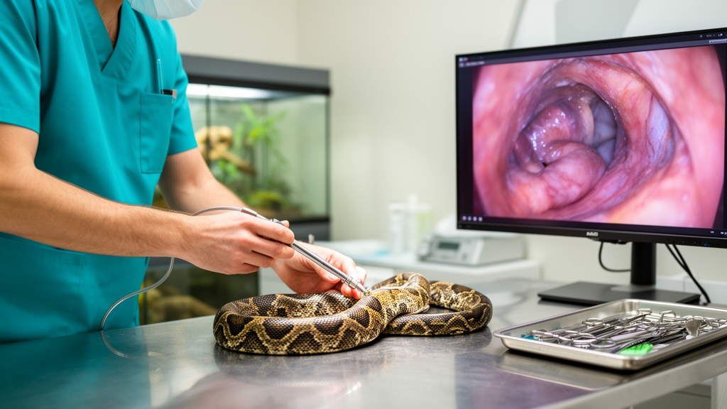
Phase 2 took place on Day 15, when each snake underwent cloacoscopic examination under pharmacological immobilization. Sedation depth and physiological parameters were monitored continuously throughout the intervention. Once sufficient muscle relaxation and immobilization were attained, each snake was positioned in dorsal recumbency and maintained in that orientation for the entire duration of the procedure. The cloacoscopic equipment comprised a 4 mm diameter, 8.5 cm long rigid endoscope with a 0° viewing angle, fitted with a protective working sheath. The endoscope was connected to an advanced imaging stack including an LED light source, a high-definition video processor, and a 26-inch monitor for real-time visualization and image recording.
Before insertion, the distal tip of the endoscope was thoroughly lubricated to facilitate atraumatic entry through the vent and prevent friction injury to the delicate cloacal mucosa. The instrument was carefully advanced through the proctodeum under direct visual guidance. The working sheath was attached to a 60 mL syringe filled with warm sterile saline solution, which was manually infused as needed throughout the examination. Rather than utilizing a fixed flow rate or preset volume, saline administration was adjusted dynamically based on luminal collapse and image clarity.
The examination revealed that cloacal architecture in all six pythonid species conforms to three distinct anatomical compartments arranged in a caudocranial sequence: the proctodeum, urodeum, and coprodeum. Because the snakes were positioned in dorsal recumbency, the investigators defined anatomical orientation based on the true dorsal and ventral surfaces of the snake rather than monitor orientation. The proctodeum formed the most caudal and narrowest chamber, largely occupied by the instrument tip upon insertion. Moving cranially, the endoscope encountered the urodeum situated dorsally and the coprodeum situated ventrally. The coprodeum communicated directly with the distal colorectal lumen via the colorectal opening, and advancing the sheath into this aperture permitted clear inspection of the distal enteric mucosa. Across all specimens, the cloacal mucosa appeared smooth, uniformly pink, and well vascularized, with no evidence of ulceration, inflammation, hemorrhage, or proliferative lesions.
Inspection of the urodeum established the principal sex-specific landmarks distinguishing male from female pythonids. In all twelve snakes, a distinct urogenital papilla was visualized in the caudodorsal region of the urodeum. In females, this papilla functioned solely as the terminal outlet of the urinary tract, whereas in males, it constituted the common outlet for both urinary and reproductive tracts. Furthermore, a transverse mucosal fold separated the urodeum from the ventral coprodeum. This fold was more prominent and clearly defined in females, whereas in males it appeared comparatively less prominent and less defined. In all female pythons, the cranial urodeum exhibited two symmetrical oviductal ostia separated by a well-developed median septum. In contrast, male pythons exhibited a complete absence of oviductal openings, with the cranial urodeum terminating as a smooth, blind-ending pouch.
The authors noted an operational distinction during the examination of male pythons: males exhibited spontaneous contractions of the cloacal musculature more frequently than females. These localized spasms caused temporary narrowing of the cloacal lumen and partial collapse of the mucosal walls, periodically obscuring the cranial recess of the urodeum. The investigators determined that this transient loss of visibility stemmed entirely from active muscular contractions rather than retained urates or fecal remnants, and infusing additional volumes of warm sterile saline readily restored luminal distension and visual clarity. Across all twelve animals, the anatomical landmarks were identified unequivocally, demonstrating that the presence of paired oviductal ostia with a median septum versus a blind-ending cranial urodeum provides an invariant morphological criterion for sex identification across the examined species.
The clinical value of direct cloacoscopy became particularly striking when comparing the endoscopic findings against the presumed sex recorded prior to the procedure. Before endoscopy, each snake had been presumptively sexed using traditional external criteria, secondary sexual traits, and manual cloacal probing. Endoscopic examination revealed that the preliminary sex assignment was incorrect in five of the twelve pythons, representing a disagreement rate of 41.7 percent. Specifically, one blood python and one Burmese python previously thought to be female were identified as males by the presence of a blind-ending urodeum without ostia. Conversely, one Burmese python, one royal python, and one reticulated python previously categorized as males were demonstrated to be females through the visualization of bilateral oviductal ostia and a median septum.
The authors emphasized that this 41.7 percent discrepancy should not be construed as a measured statistical error rate for cloacal probing, nor does it establish formal diagnostic superiority, because the study was descriptive rather than a blinded, controlled diagnostic accuracy trial. Nonetheless, the high discrepancy underscores the practical limitations of conventional indirect sexing methods in juvenile and subadult snakes.
Beyond sex determination, the authors highlighted that cloacoscopy provides a practical platform for broader clinical and reproductive interventions. Furthermore, direct identification of the female reproductive openings may facilitate controlled access to the reproductive tract and holds potential for assisted reproductive procedures, including artificial insemination, which has previously been applied in snakes. Future studies evaluating neonatal pythons immediately after hatching will be necessary to determine whether these urodeum landmarks are sufficiently developed at birth to allow neonatal sexing, which would offer invaluable support to global reptile breeding and conservation management.
Subject of Research: Endoscopic cloacal anatomy and sex determination in captive Pythonidae
Article Title: Endoscopic findings on cloacal anatomy and sex identification in Pythonidae
Article References: Matteo, M., Stella, C., Marco, D. G., & Filippo, S. (2026). Endoscopic findings on cloacal anatomy and sex identification in Pythonidae. Frontiers in Veterinary Science, 13, Article 1924663. https://doi.org/10.3389/fvets.2026.1924663
Image Credits: AI Generated
DOI: 10.3389/fvets.2026.1924663
Keywords: Pythonidae, cloacoscopy, endoscopy, sex determination, veterinary medicine, herpetology, urodeum, proctodeum, coprodeum, oviductal ostia, alfaxalone, reptile reproduction, assisted reproduction, Python molurus bivittatus, Python brongersmai, Morelia spilota, Malayopython reticulatus, Python regius, Python sebae
News Source: Drew Townsend. (November 11, 2026). Cloacoscopy Reveals Sex-Specific Anatomical Structures in Diverse Captive Python Species. Scienmag.