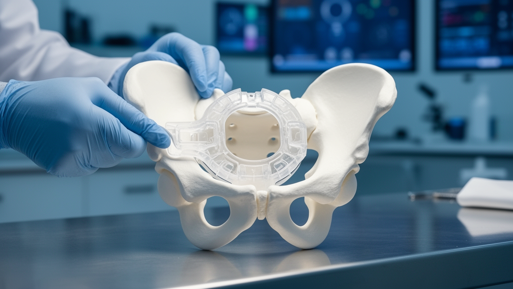
Orthopedic surgeons have successfully utilized digital simulation and a patient-specific three-dimensional printed osteotomy guide to correct a severe, multiplanar pediatric hip deformity in a ten-year-old patient. The clinical case, documented by researchers from Guizhou University of Traditional Chinese Medicine and Beijing Jishuitan Hospital Guizhou Hospital, illustrates how preoperative virtual modeling can translate complex spatial planning into direct intraoperative execution for conditions where standard surgical approaches face substantial geometric challenges.
The patient presented with a five-year history of persistent right hip pain and progressive restriction of joint mobility. Clinical physical examination revealed pronounced functional deficits, including approximately four centimeters of right-sided leg-length discrepancy, proximal migration of the right greater trochanter, and marked limitation of hip motion across multiple anatomical planes. In older pediatric populations, the intrinsic capacity for spontaneous bone remodeling is notably diminished, heightening the necessity for precise structural realignment during primary surgical intervention.
Comprehensive diagnostic imaging established a compound structural pathology involving several coexisting abnormalities. Bilateral hip digital radiography, three-dimensional computed tomography reconstruction, and magnetic resonance imaging demonstrated right coxa vara accompanied by femoral head retroversion, an old femoral neck fracture exhibiting possible nonunion, suspected avascular necrosis of the femoral head, and significant limb shortening. In such multiplanar presentations, conventional surgical planning based solely on two-dimensional radiographs and intraoperative visual estimation can prove insufficient to accurately calculate deformity angles and determine optimal fixation trajectories.
To overcome these anatomical complexities, the surgical team imported the patient’s preoperative computed tomography data into Mimics software to generate a one-to-one three-dimensional digital reconstruction. Through this virtual environment, the surgeons quantified the multiplanar skeletal distortions and performed virtual osteotomy planning. The simulated spatial parameters were subsequently used to design and manufacture a customized, patient-specific three-dimensional printed osteotomy guide intended to direct intraoperative localization, bone cuts, and hardware placement.
During the operative procedure, the customized printed guide assisted the surgeons in performing a targeted proximal femoral osteotomy, multiplanar deformity correction, rigid plate-and-screw internal fixation, and structural bone grafting directly at the osteotomy interface. The intervention was complemented by postoperative hip bracing. The entire surgical procedure required three hours and forty minutes of operative time, incurred an estimated blood loss of 200 milliliters, and utilized intraoperative C-arm fluoroscopy on three occasions to confirm alignment and implant positioning.
Short-term clinical follow-up at four months demonstrated substantial symptomatic and functional recovery. The patient reported complete relief of right hip pain and displayed marked improvements in ambulatory gait mechanics. Orthopedic re-examination confirmed that the right-sided limb shortening had decreased from approximately four centimeters down to less than one centimeter. Follow-up diagnostic imaging confirmed satisfactory bone healing at the osteotomy site, maintenance of stable internal fixation, and restored congruence between the femoral head and the acetabulum.
Despite the successful outcome, the study authors emphasize that findings derived from an individual case report cannot definitively demonstrate superior surgical precision, reduced radiation exposure, or clear advantages over traditional reconstructive techniques. The investigators conclude that prospective comparative investigations involving larger patient cohorts and extended follow-up periods remain necessary to systematically evaluate guide-fit error margins, operative accuracy, radiation exposure, cost-effectiveness, and the long-term maintenance of anatomical correction throughout ongoing skeletal growth.
Subject of Research: Digital simulation and 3D-printed guide assistance in pediatric hip deformity surgery
Article Title: Digital simulation and 3D-printed guide-assisted surgical correction of a complex pediatric hip deformity: a case report and literature review
Article References: Zeng, M., Wu, S., & Mo, A. (2026). Digital simulation and 3D-printed guide-assisted surgical correction of a complex pediatric hip deformity: a case report and literature review. Frontiers in Pediatrics, 14, Article 1991427. https://doi.org/10.3389/fped.2026.1991427
Image Credits: AI Generated
DOI: 10.3389/fped.2026.1991427
Keywords: pediatric orthopedics, 3D printing, digital simulation, proximal femoral osteotomy, coxa vara, femoral head retroversion, femoral neck fracture, avascular necrosis, patient-specific guide, virtual surgical planning, Mimics software, leg-length discrepancy, plate-and-screw fixation, bone grafting, hip bracing, fluoroscopy, case report
News Source: Harold Sullivan. (November 11, 2026). Custom 3D-Printed Guide Aids Pediatric Hip Deformity Correction in Case Study. Scienmag.