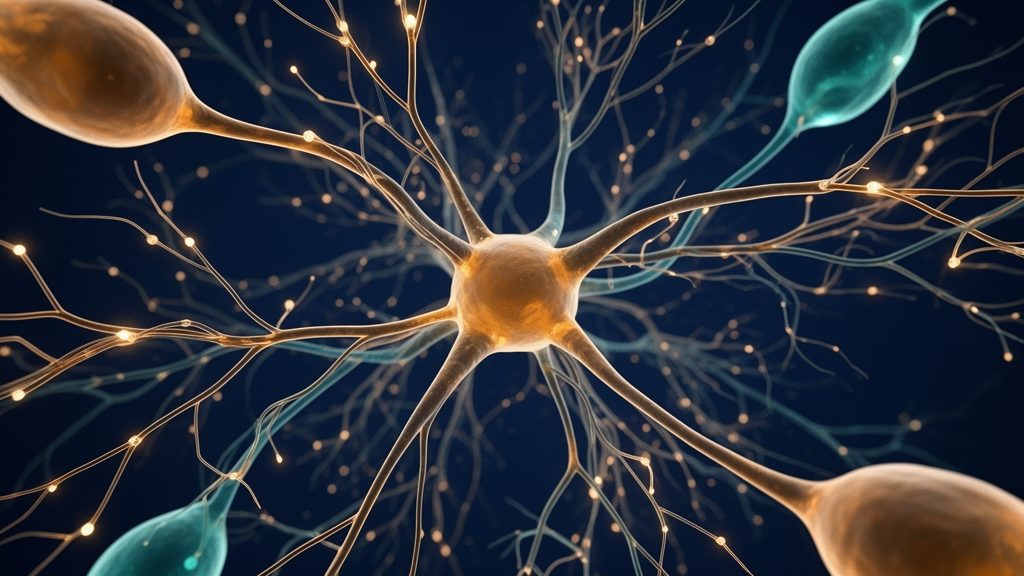
In an experimental study published in Current Biology, researchers at the Max Planck Florida Institute for Neuroscience demonstrated that neurons in the primary visual cortex of ferrets refine their circuitry immediately following the onset of visual experience primarily by adding vast numbers of new excitatory connections rather than pruning unneeded inputs. Investigating how early sensory input reshapes the brain, the team discovered that cortical neurons expand their dendritic spines nearly five-fold during the initial days after eye opening, systematically organizing these new synaptic inputs to sharpen responses to visual stimuli.
For decades, neuroscientists have largely held that early circuit refinement occurs predominantly through pruning, a regressive developmental process in which the brain selectively preserves functional pathways while eliminating redundant or poorly matched connections. However, the new findings from the laboratory of Dr. David Fitzpatrick reveal that this classical model omits an essential constructive phase.
Prior to eye opening, genetic programs establish an initial first draft of the brain’s visual architecture, constructing a basic neural scaffold capable of receiving signals. At this nascent stage, neurons within the primary visual cortex respond broadly across diverse visual features, and the inputs arriving from the two eyes do not yet match. Under normal development, the introduction of visual experience takes control over the system, guiding the circuit so that individual neurons become sharply tuned to specific stimulus features and the separate signals arriving from both eyes become properly aligned for mature vision.
To capture the cellular dynamics underlying this transformation, co-lead authors Dr. Juliane Jaepel and Dr. Clara Tepohl mapped the individual synaptic inputs received by single neurons at the exact onset of eye opening and again after several days of natural visual experience. Synaptic connections from other neurons terminate at dendritic spines, microscopic protrusions extending along a neuron’s dendritic arbor.
During these mapping experiments, the researchers presented controlled visual patterns to each eye independently. This testing paradigm allowed them to assess whether an individual dendritic spine responded selectively to the left eye, the right eye, or both eyes simultaneously. In addition, the scientists determined which specific visual features—such as edge orientation—evoked the strongest physiological responses at each spine, and tracked whether spines responsive to similar visual features were positioned adjacent to one another along the dendritic branches.
Quantitative comparisons between maps obtained at eye opening and those recorded after days of visual experience demonstrated that the total population of dendritic spines expanded nearly five-fold. Rather than representing an indiscriminate proliferation of synapses, this expansion exhibited pronounced functional specificity. Excitatory connections tuned selectively to edges of a particular orientation—a fundamental building block of spatial vision—expanded substantially more than non-selective connections that responded equally to all orientations.
The study also revealed a marked structural reorganization in the binocular inputs received by cortical neurons. Following the onset of visual experience, the number of dendritic spines showing matched selectivity to the same edge orientation across both eyes increased dramatically.
Beyond altering the composition and tuning of synaptic inputs, visual experience also dictated the spatial arrangement of connections along the dendritic branches. After early visual exposure, dendritic spines that responded to similar edge orientations were frequently localized immediately adjacent to one another.
Senior author Dr. David Fitzpatrick noted that as visual experience commences, neurons reorganize their network of synaptic connections by building directly upon the pre-existing genetic scaffold, tailoring the specific patterns of inputs they receive to support more robust and coherent sensory processing. Co-lead author Dr. Juliane Jaepel highlighted that the nearly five-fold increase in excitatory connections directly drives the dramatic functional remodeling that converts broadly tuned juvenile circuits into mature, feature-selective networks.
The research team suggested that this rapid, expansion-based rewiring may reflect a universal principle of neural development that operates across sensory, cognitive, and behavioral circuits shaped by early life experience. Supported by the National Institutes of Health, the Max Planck Florida Institute for Neuroscience, and the Max Planck Society, future investigations into the cellular mechanisms controlling early circuit growth could provide valuable insights into how disruptions in early synaptic formation contribute to developmental brain disorders.
Reconstructions of example neurons in the primary visual cortex illustrate the structural trajectory of circuit remodeling between the initial opening of the eyes and the subsequent two to three weeks of visual experience. In these anatomical reconstructions, where scale is indicated by a 50-micron scale bar, each individual dot marks a dendritic spine that serves as the physical site of an excitatory connection received from another neuron. Over this two-to-three-week window of sensory maturation, cortical neurons physically grow larger and accumulate substantially greater numbers of synaptic connections across their branches.
Subject of Research: Experience-dependent synaptic addition and functional circuit refinement in the ferret primary visual cortex
Article Title: MPFI researchers uncover new insights into circuit rewiring following the onset of visual experience
Article References: MPFI researchers uncover new insights into circuit rewiring following the onset of visual experience. Original publication (Bibliographic details unavailable.)
Image Credits: AI Generated
DOI: Not provided
Keywords: primary visual cortex, visual experience, dendritic spines, synaptic plasticity, circuit rewiring, Max Planck Florida Institute, Current Biology, David Fitzpatrick, Juliane Jaepel, Clara Tepohl, edge orientation, binocular alignment, ferret visual system, excitatory synapses, neural development, synaptic clustering, cortical circuits
News Source: Drew Townsend. (November 11, 2026). Ferret Visual Cortical Neurons Rapidly Multiply Connections Following Initial Visual Experience. Scienmag.