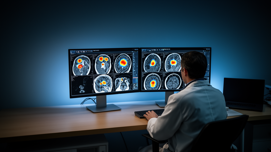
A prospective clinical evaluation has demonstrated that applying specialized deep learning models to digital positron emission tomography scanners built with bismuth germanate detectors enables diagnostic scans in sixty to ninety seconds while preserving lesion conspicuity and noise suppression. The study investigated a new generation of digital positron emission tomography combined with computed tomography systems that employ long axial fields of view and bismuth germanate scintillation crystals paired with silicon photomultipliers. Although bismuth germanate crystals provide high linear attenuation coefficients for five hundred and eleven kiloelectronvolt annihilation photons, lack intrinsic radioactivity, and cost substantially less than traditional lutetium-based scintillators, their lower light yields and prolonged scintillation decay times preclude hardware-based time-of-flight measurement. To overcome this limitation, medical physicists evaluated deep learning architectures designed to emulate time-of-flight information on non-time-of-flight acquisition data, establishing that combining sixty to ninety second scan times with penalization factors between three hundred and five hundred under a medium-strength neural model delivers optimal image performance.
Positron emission tomography serves as an indispensable molecular imaging modality across clinical oncology, neurology, cardiology, and the evaluation of complex inflammatory disorders by quantifying cellular biochemical processes at the molecular scale. The introduction of deep learning-based time-of-flight algorithms known as Precision Deep Learning on the Omni Legend system seeks to restore these spatial localization benefits mathematically. These algorithms operate downstream from block-sequential regularized expectation maximization, an advanced iterative reconstruction technique incorporating a relative difference prior to ensure absolute voxel-wise mathematical convergence while suppressing background noise via a single global regularization parameter denoted as beta.
To rigorously delineate the mathematical boundaries and clinical performance of these combined computational tools, researchers designed a prospective protocol encompassing twenty consecutive oncology patients referred for routine clinical examinations. Each participating subject gave written informed consent under regional ethical board approval conforming to the Declaration of Helsinki. To ensure sufficient lesion visibility across experimental reconstructions, the inclusion criteria required each patient to exhibit at least two visually identifiable fluorodeoxyglucose-avid lesions on standard clinical reference scans acquired at ninety seconds per bed position with a beta penalization setting of four hundred. Each participant received an intravenous administration of fluorine-18 fluorodeoxyglucose standardized precisely to three point five megabecquerels per kilogram of body mass. Scans were initiated sixty minutes after radiotracer injection on a digital scanner featuring a thirty-two centimeter axial field of view, acquiring list-mode emission data for one hundred and twenty seconds per bed position spanning from the base of the skull through the mid-thigh level.
The raw list-mode emission streams were systematically rebinned into distinct temporal durations to emulate different clinical acquisition protocols, creating independent datasets corresponding to one hundred and twenty, ninety, sixty, forty-five, thirty, and fifteen seconds per bed position. Each temporal dataset was then reconstructed into three hundred and eighty-four by three hundred and eighty-four voxel matrices using block-sequential regularized expectation maximization across seven discrete beta penalization factors spanning fifty, one hundred, three hundred, five hundred, seven hundred, nine hundred, and eleven hundred. These reconstructions were subsequently processed through three distinct deep learning time-of-flight configurations designated as Low, Medium, and High models, which were initially trained on paired non-time-of-flight and hardware time-of-flight patient images reconstructed with block-sequential regularization. For clinical comparison, investigators also generated baseline reference reconstructions from the ninety-second acquisitions using conventional ordered subset expectation maximization with three iterations, twenty-two subsets, point spread function modeling, and a three-millimeter post-reconstruction Gaussian filter.
Quantitative assessment required robust and objective metrics capable of isolating signal quality from anatomical variability across diverse organ systems. Across the twenty patients, investigators evaluated forty target lesions with an average volume of one point two milliliters, ranging from zero point one to three point seven milliliters, distributed across the pelvic region, abdomen, thorax, and head and neck. Spherical volumes of interest fifty millimeters in diameter were placed in the upper portion of the right hepatic lobe to measure healthy liver uptake, alongside dedicated volumes of interest positioned over each avid tumor. To avoid the known measurement instability of maximum standardized uptake values, which isolate only the single highest-intensity voxel and remain highly vulnerable to statistical image noise, the researchers extracted peak standardized uptake values defined as the mean uptake within a one-milliliter spherical volume enclosing the highest local activity. Signal-to-noise ratios were calculated as mean liver uptake divided by liver standard deviation, while lesion detectability was quantified through a normalized peak-to-peak ratio comparing lesion peak uptake directly against liver peak uptake.
Experimental measurements demonstrated that liver signal-to-noise ratios increased consistently with prolonged acquisition durations and higher beta penalization factors across every reconstruction permutation, ranging from a minimum of zero point seven for fifteen-second acquisitions reconstructed at beta fifty up to twenty-four point three for two-minute scans reconstructed at beta eleven hundred. Incorporating neural models consistently elevated signal-to-noise performance, with the Low deep learning model yielding the most pronounced noise suppression, achieving up to a one hundred and four percent improvement in signal-to-noise ratio over conventional reconstructions at one hundred and twenty seconds and beta fifty. Conversely, the High deep learning model produced a less pronounced increase across increasing beta values and caused a twenty-three percent reduction in signal-to-noise ratio at beta eleven hundred during two-minute acquisitions. This divergence reflects the underlying neural training objectives, wherein the Low model was parameterized to emulate high-beta target images prioritizing aggressive noise filtering, while the High model targeted low-beta profiles prioritizing structural contrast retention.
Analysis of lesion metrics revealed that tumor peak standardized uptake values remained largely independent of acquisition duration across all evaluated durations. However, peak uptake within tumors decreased steadily as beta values increased across unenhanced reconstructions as well as those processed with Low and Medium deep learning models, illustrating the expected edge-smoothing effects of high mathematical regularization. In contrast, when the High deep learning model was applied to acquisitions of sixty seconds or longer, lesion peak values remained remarkably stable across beta penalizations from five hundred to eleven hundred. The High neural model produced the highest overall peak tumor values relative to the standard ninety-second ordered subset expectation maximization baseline, demonstrating that contrast-focused neural processing counteracts the loss of peak intensity typically induced by heavy regularizing priors during iterative convergence.
Lesion detectability, governed by the peak-to-peak ratio between tumor uptake and background liver uptake, exhibited complex dependencies on both scan duration and regularization strength. For unassisted reconstructions at extended acquisition durations of ninety and one hundred and twenty seconds, the greatest detectability occurred at a low penalization value of beta one hundred, indicating that minimal smoothing is required when photon count statistics are abundant. When scan durations were compressed to sixty seconds or less, higher regularization at beta three hundred became necessary to suppress elevated statistical noise and maintain lesion conspicuity. Across the neural networks, the High model generally yielded superior detectability across experimental settings, peaking at beta nine hundred for longer scan times, whereas the Low and Medium models peaked at lower penalization values between beta three hundred and five hundred. The Low model achieved optimal noise suppression but did so at the direct expense of lesion detectability, confirming that aggressive noise smoothing blunts diagnostic peak contrast.
To verify that these quantitative clinical relationships reflected true physical imaging phenomena rather than regional anatomical variations or patient-specific physiological fluctuations, the authors conducted validation trials using an anthropomorphic image quality thorax phantom. The experimental phantom incorporated a fillable zero point five milliliter spherical insert configured with a four-to-one sphere-to-background radioactivity ratio, immersed within a uniform background activity concentration of four point eight kilobecquerels per milliliter of fluorine-18 representing typical hepatic biodistribution during clinical examinations. Five repeated list-mode acquisitions of two minutes each were obtained with the sphere centered within the axial field of view, followed by identical rebinning, iterative reconstruction, and deep learning post-processing. Quantitative measurements derived from the phantom verified the exact trends observed in the clinical cohort, reproducing the characteristic trade-offs between signal-to-noise gains and peak contrast preservation across identical combinations of beta regularization and neural model strength.
The study authors emphasized critical clinical boundaries, observing that no degree of algorithmic reconstruction or artificial intelligence enhancement can synthesize or recover diagnostic lesions if sufficient photon counts are absent from the primary projection data, as demonstrated in thirty-second clinical acquisitions where small lesions remained entirely undetectable across all models. Furthermore, the findings reflect a fixed injected dosage of three point five megabecquerels per kilogram administered to predominantly lean individuals presenting with established avid lesions, meaning performance in heavier patients or lower-dose protocols warrants continued empirical validation. Because the peak-to-peak ratio serves as a physical surrogate metric rather than direct human diagnostic efficacy, comprehensive reader studies will be required before clinical adoption. Nevertheless, for digital bismuth germanate scanners operating under standard fluorodeoxyglucose protocols, pairing sixty to ninety second acquisitions with beta factors of three hundred to five hundred under medium neural post-processing establishes an optimal operational standard.
Subject of Research: Evaluation of deep learning time-of-flight reconstruction models for digital non-TOF BGO PET/CT imaging
Article Title: Evaluation of deep learning-based reconstruction models on non-TOF BGO PET/CT: impact of acquisition times and BSREM penalization factors on lesion detectability and SNR
Article References: Stenvall, A., Minarik, D., Trägårdh, E., & Kvernby, S. (2026). Evaluation of deep learning-based reconstruction models on non-TOF BGO PET/CT: impact of acquisition times and BSREM penalization factors on lesion detectability and SNR. EJNMMI Physics, 13(1), Article 74. https://doi.org/10.1186/s40658-026-00919-8
Image Credits: AI Generated
DOI: 10.1186/s40658-026-00919-8
Keywords: Positron Emission Tomography, Bismuth Germanate Detectors, Deep Learning Reconstruction, Time of Flight PET, Fluorodeoxyglucose, BSREM Reconstruction, Signal to Noise Ratio, Lesion Detectability, Digital PET-CT, Standardized Uptake Value, Penalization Factor, Image Quality Optimization, Medical Image Processing, Anthropomorphic Phantom, Silicon Photomultipliers, Radiation Count Statistics, Clinical Protocol Optimization
News Source: Blake Davidson. (November 11, 2026). Deep Learning Enhances Tumor Detectability in Fast Non-TOF BGO PET Scans. Scienmag.