A single-case longitudinal study from a Nigerian tertiary hospital shows that a ten-week multimodal physiotherapy program achieved significant functional recovery and pain reduction in a patient with rotator cuff tendinitis and subacromial bursitis following a corticosteroid injection. Rotator cuff tendinitis and subacromial bursitis are prevalent causes of shoulder pain and disability, yet corticosteroid injections frequently provide only temporary relief without resolving underlying neuromuscular deficits. In this report, a seventy-one-year-old female patient presented with severe right shoulder impairment, recording a baseline score of seven out of ten on the Numerical Pain Rating Scale and a disability rating of 67.7 percent on the Shoulder Pain and Disability Index. Following the ten-week multimodal intervention, her worst pain decreased to two out of ten, reflecting an absolute reduction of five points, while her disability rating dropped to 16.3 percent, demonstrating an absolute improvement of 51.4 percent. Both outcomes substantially exceeded established minimal clinically important difference thresholds, demonstrating that structured physical therapy plays an essential role in securing sustained functional recovery after corticosteroid administration.
The clinical presentation involved a seventy-one-year-old retired female nurse who sought care after experiencing sudden-onset right shoulder pain approximately three weeks prior to presentation, with no preceding trauma or fall. The patient described the pain as an intermittent, aching sensation localized to the anterolateral shoulder with radiation down the arm. Symptoms were present at rest and worsened substantially during overhead movements and lifting, with nocturnal pain repeatedly interrupting her sleep. Her medical history included peptic ulcer disease, and past drug therapy included a right shoulder corticosteroid injection, celecoxib, and omeprazole, though she was taking no regular analgesics upon assessment. She had no past surgical history. Family and social evaluations recorded that she lived with her partner in a bungalow, was married in a monogamous arrangement with four children, and abstained from tobacco and alcohol. Functional restrictions prevented her from lifting objects with the right upper extremity or performing household chores, forcing reliance on the unaffected left arm for daily living activities.
Initial physical examination confirmed that the patient entered the clinic unaided, walking with a normal gait and symmetrical arm swing. She was afebrile, acyanosed, anicteric, and in no respiratory distress, although mild swelling was observable over the anterolateral right shoulder, with intact skin and no discoloration. A complete review of systems demonstrated preserved neurological and cognitive status, intact sensation, normal limb reflexes, and unimpaired balance without hypertonia or spasticity. Baseline vital signs remained within normal physiological ranges, recording a blood pressure of 110/70 mmHg, a pulse rate of 72 beats per minute, a respiratory rate of 23 cycles per minute, and a body temperature of 37 degrees Celsius. Auscultation confirmed clear vesicular breath sounds with symmetrical chest motion, while distal examinations identified no peripheral edema in the hands or wrists. The integumentary system exhibited pliable, hydrated skin without trophic abnormalities or temperature asymmetry, and gastrointestinal and urinary continence were intact.
Musculoskeletal assessment at baseline documented severe right shoulder capsular tightness, localized tenderness over the subacromial space and greater tuberosity, and preserved overall muscle bulk. Active range of motion testing in the right shoulder elicited a pain rating of seven out of ten on the Numerical Pain Rating Scale, whereas passive range of motion testing provoked an aggravated pain response of nine out of ten. Manual muscle evaluation using the Oxford grading scale revealed marked weakness isolated to the affected right shoulder girdle, showing muscle strength grades of three out of five for shoulder flexors, extensors, abductors, and internal rotators, alongside four out of five for adductors and external rotators. Distal joint musculature demonstrated intact strength, with right elbow flexors, elbow extensors, wrist flexors, and wrist extensors all grading five out of five. Furthermore, all muscle groups within the unaffected left upper extremity and both lower extremities demonstrated full five-out-of-five strength, confirming that functional deficits were confined entirely to the right shoulder complex.
Physical diagnostic maneuvers corroborated local rotator cuff and subacromial pathology while ruling out cervical root involvement and full-thickness tears. Positive provocative signs included the painful arc test, with pain localized at 90 degrees and 170 degrees of abduction, along with positive results for Hawkins-Kennedy, Speed, sulcus, empty can, and resisted isometric internal rotation and abduction tests. In contrast, Spurling’s test, the cervical compression test, and the drop arm test were negative. Diagnostic imaging supported the clinical evaluation. Plain radiography of the right shoulder revealed degenerative changes at the acromioclavicular joint, sclerotic alterations at the acromion and distal third of the clavicle, and osteopenia of the right clavicle. Soft-tissue ultrasound examination demonstrated cortical irregularity of the greater tuberosity and a moderate hypoechoic effusion surrounding the long head of the biceps tendon. Importantly, ultrasound confirmed normal morphology across the supraspinatus, infraspinatus, teres minor, and subscapularis tendons without partial or full-thickness tears, confirming a diagnosis of tendinitis affecting the supraspinatus and subscapularis muscles with associated subacromial bursitis.
The clinical profile reflected broader medical understanding regarding rotator cuff disorders, commonly termed shoulder impingement syndrome or subacromial pain syndrome. This disorder involves tendon inflammation, microvascular degeneration, and localized pain within the rotator cuff musculature—primarily the supraspinatus, though frequently engaging the subscapularis and infraspinatus—frequently coexisting with subacromial bursitis. The etiology encompasses intrinsic tendon degeneration, including hypovascularity and disorganized collagen architecture that diminish tensile capacity, combined with extrinsic compression from osteophytes, acromial variations, scapular dyskinesis, and repetitive overhead tasks. Globally, shoulder pain impacts between 7 percent and 34 percent of the general population, representing a leading source of disability in adults aged over forty. In Nigeria, epidemiological data from a tertiary hospital in the south-west indicates that shoulder disorders represent a substantial fraction of outpatient physical therapy cases, predominantly affecting older adults and manual workers. While conservative non-surgical management achieves favorable recovery in approximately 70 to 80 percent of patients within six to twelve weeks, unaddressed pathology risks chronic tears, adhesive capsulitis, and functional disability.
To overcome these biomechanical and inflammatory deficits, the clinical team designed a structured ten-week multimodal rehabilitation regimen paired with a daily home exercise program of pendulums, active-assisted range of motion, and isometric loading. Addressing the acute to subacute inflammatory presentation, physiotherapists selected cryotherapy rather than thermotherapy during the initial phase. Cryotherapy provided targeted vasoconstriction, decreased cellular metabolic demand, and slowed sensory nerve conduction velocity, effectively producing analgesia and limiting exercise-induced symptom flare-ups without exacerbating edema. Conversely, superficial heat was contraindicated early due to the risk of aggravating swelling in inflamed tissues. Therapeutic ultrasound was incorporated as an adjunct during initial care to support pain modulation and tissue heating, improving the patient’s capacity to engage in active rehabilitation. The report acknowledged, however, that clinical evidence supporting ultrasound as an isolated modality remains inconsistent, leading practitioners to position it as a secondary adjunct alongside evidence-backed active movement therapies.
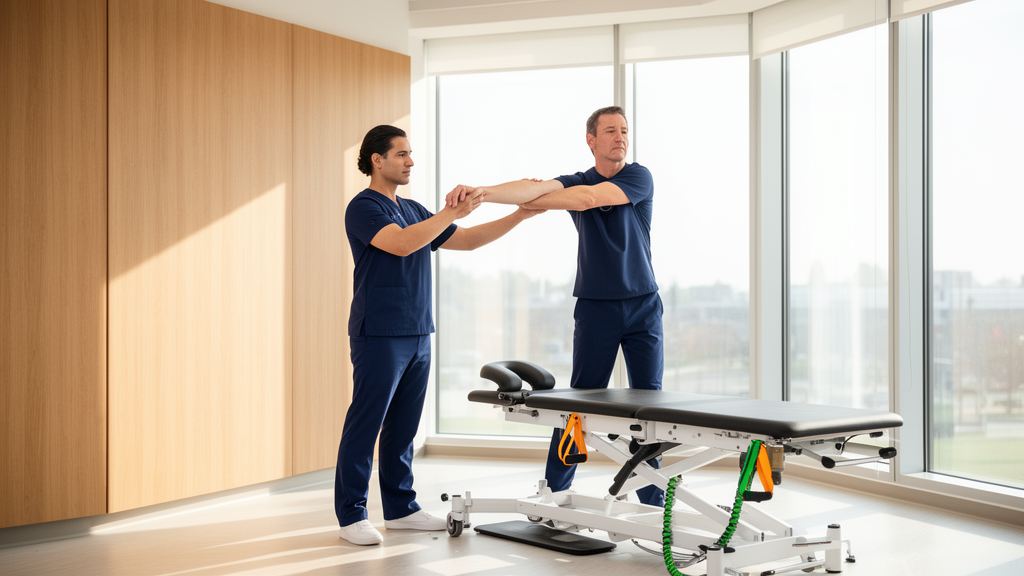
As the acute inflammatory phase stabilized, the therapeutic focus shifted toward progressive loading, joint mobilization, and kinetic chain re-education. Eccentric strengthening exercises were integrated to stimulate tendon remodeling, enhance load tolerance, and alleviate persistent tendinopathic pain. Concurrently, manual therapy techniques involving glenohumeral joint mobilizations and myofascial soft tissue release were utilized to reduce capsular tightness, restore normal arthrokinematics, and modulate peripheral pain signals. Targeted strengthening of scapular stabilizing musculature, particularly the serratus anterior and lower trapezius, was systematically applied to restore proper scapulothoracic rhythm during arm elevation, thereby reducing abnormal mechanical compression against the subacromial space. Alongside supervised clinic sessions, comprehensive patient education addressing load management, postural awareness, and ergonomic modifications was emphasized. This educational component aimed to foster self-management strategies, facilitate independent execution of daily home exercises, and minimize the risk of mechanical reinjury during household activities.
Post-intervention evaluations following the ten-week program documented significant quantitative improvements that exceeded established minimal clinically important difference thresholds across all primary outcome domains. Pain intensity decreased by 75 percent at rest and 71.4 percent during movement, with worst pain ratings dropping from seven out of ten to two out of ten, surpassing the two-point threshold on the Numerical Pain Rating Scale. The patient’s functional disability score on the Shoulder Pain and Disability Index improved from 67.7 percent to 16.3 percent, representing a 75.9 percent relative improvement and a 51.4 percent absolute reduction that exceeded the clinical threshold of 8 to 13 points. Active range of motion expanded beyond the standard ten-degree threshold in all cardinal planes: shoulder flexion increased from 110 to 168 degrees, abduction rose from 85 to 166 degrees, external rotation improved from 40 to 75 degrees, and internal rotation advanced from 37 to 75 degrees, with notable gains in extension and adduction. Shoulder muscle strength improved by one Oxford grade across all tested groups, and nocturnal pain resolved, restoring comfortable sleep on the affected side.
Despite these positive functional gains, the study authors highlighted key methodological constraints associated with their single-case longitudinal design. Evaluating an individual patient in a single clinical environment without a comparative control group restricts the direct generalizability of the findings across wider demographic populations. In accordance with methodological standards for single-subject research, outcomes were assessed via descriptive statistics, absolute change scores, and percentage improvements rather than inferential statistical testing. The authors noted that larger randomized controlled trials are required to rigorously evaluate multimodal physiotherapy protocols and assess adjunctive modalities like therapeutic ultrasound against alternative interventions. Additionally, long-term longitudinal investigations are needed to evaluate the durability of functional gains, monitor patient quality of life, and measure rates of potential progression toward surgical repair. Future investigations incorporating qualitative methodologies, such as structured patient interviews, could also provide valuable insights into adherence barriers, thereby refining personalized rehabilitation strategies for rotator cuff pathologies.
Subject of Research: Multimodal physiotherapy management of rotator cuff tendinitis and subacromial bursitis
Article Title: Physiotherapy management of rotator cuff tendinitis with associated subacromial bursitis in a tertiary hospital in Nigeria: a case report
Article References: Agunbiade, J. T., Bello, R. A., Oshomoji, O. I., Ogungbe, O. A., & Echebi, L. U. (2026). Physiotherapy management of rotator cuff tendinitis with associated subacromial bursitis in a tertiary hospital in Nigeria: a case report. Bulletin of Faculty of Physical Therapy, 31(1), Article 1. https://doi.org/10.1186/s43161-025-00336-5
Image Credits: AI Generated
DOI: 10.1186/s43161-025-00336-5
Keywords: Rotator cuff tendinitis, Subacromial bursitis, Physical therapy, Shoulder rehabilitation, Corticosteroid injection, SPADI, Musculoskeletal disorders, Case report, Physiotherapy, management, rotator, cuff
News Source: Ophelia Keating. (November 10, 2026). Multimodal Physiotherapy Restores Shoulder Mobility Following Corticosteroid Injection in Case Study. Scienmag.