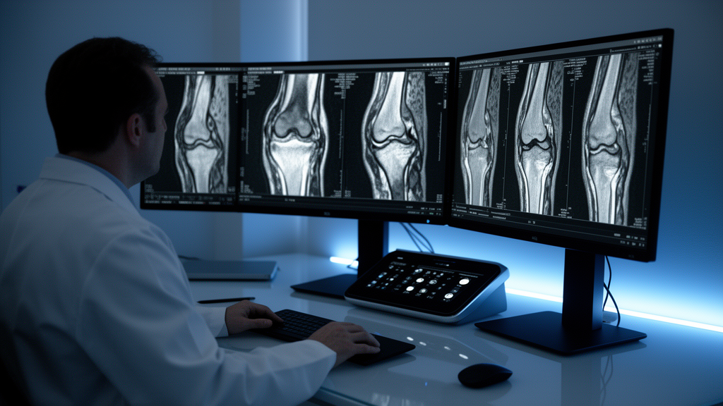
Complex tears represented the most common pattern of knee joint meniscal tears in a retrospective cross-sectional evaluation of patients evaluated with magnetic resonance imaging at a diagnostic facility in Kano State, Nigeria. Researchers Fatima Abba Mukhtar, Maryam O. Hassan, and Possible W. Ewona reported that complex tears accounted for 34.0 percent of observed tear patterns among the reviewed cohort, followed by horizontal tears at 26.0 percent and bucket-handle tears at 14.0 percent. The investigation, published in BMC Musculoskeletal Disorders, focused on establishing a baseline of imaging-diagnosed tear characteristics and associated joint abnormalities in a region where musculoskeletal magnetic resonance imaging evidence has remained limited.
Meniscal tears serve as a frequent cause of knee pain and physical disability, with magnetic resonance imaging functioning as the principal noninvasive imaging modality employed to evaluate suspected tears of the meniscus. Although magnetic resonance imaging is widely used for this diagnostic purpose, its documented diagnostic performance and the specific patterns of identified tears vary substantially across different clinical environments. The researchers noted that diagnostic data regarding knee injuries from northern Nigeria have been sparse, motivating the review of localized imaging presentations to describe tear morphology, precise anatomical locations, and concurrent joint pathologies.
The investigation utilized a descriptive, retrospective cross-sectional study design conducted at the Medserve Kano Diagnostic Centre in Kano, Nigeria. The research team examined archived magnetic resonance images and corresponding radiology reports for 50 patients who underwent knee magnetic resonance imaging for suspected meniscal pathology between July 2025 and February 2026. All patient examinations were performed using a 1.5-Tesla Siemens scanner. Formal ethical approval for conducting the archival review was granted by the Kano State Ministry of Health Research Ethics Committee prior to data analysis.
Among the 50 reviewed patients, male participants formed the vast majority of the study cohort, comprising 43 individuals or 86.0 percent of the total sample, while female participants accounted for 7 individuals or 14.0 percent. In evaluating the specific anatomical distribution of tears among participants with recorded meniscal horn locations, involvement of the posterior horn emerged as the most frequently identified single site. Posterior horn tears were documented in 63.0 percent of recorded medial meniscal tears and in 37.9 percent of recorded lateral meniscal tears.
The review revealed that nine individuals, representing 18.0 percent of the study participants, exhibited no definable tear pattern and had no magnetic resonance imaging-verified tear in either the medial or lateral meniscus, despite their clinical referral for suspected meniscal injury. These participants nonetheless demonstrated other concurrent joint abnormalities on their scans. Across the broader study group, documented associated magnetic resonance imaging findings included anterior cruciate ligament injuries, bone bruises, osteoarthritis-related joint changes, and joint effusions.
Data were processed using descriptive statistics in SPSS version 26.0, alongside exploratory Fisher’s exact tests applied to evaluate selected categorical comparisons. These exploratory analyses yielded no statistically verifiable evidence of associations between patient age group and tear complexity, patient sex and tear complexity, or anterior cruciate ligament injury status and meniscal tear location. The authors emphasized that because the study relied on a limited sample size yielding wide confidence intervals, all exploratory statistical findings must be interpreted with caution. They concluded that further multicenter research involving larger cohorts and correlation with arthroscopic findings is necessary to validate these descriptive baseline observations.
Subject of Research: Magnetic resonance imaging evaluation of knee joint meniscal tears in Kano State, Nigeria
Article Title: Magnetic resonance imaging evaluation of knee joint meniscal tears in a selected diagnostic centre in Kano state, Nigeria
Article References: Mukhtar, F. A., Hassan, M. O., & Ewona, P. W. (2026). Magnetic resonance imaging evaluation of knee joint meniscal tears in a selected diagnostic centre in Kano state, Nigeria. BMC Musculoskeletal Disorders. https://doi.org/10.1186/s12891-026-10702-6
Image Credits: AI Generated
DOI: 10.1186/s12891-026-10702-6
Keywords: knee joint, meniscal tears, magnetic resonance imaging, 1.5-T Siemens scanner, complex tears, horizontal tears, bucket-handle tears, posterior horn, medial meniscus, lateral meniscus, anterior cruciate ligament, bone bruise, osteoarthritis, joint effusion, Medserve Kano Diagnostic Centre, Kano State, Nigeria, retrospective cross-sectional study
News Source: Ophelia Keating. (November 10, 2026). Complex Knee Meniscal Tears Predominate in Kano Diagnostic Centre MRI Study. Scienmag.