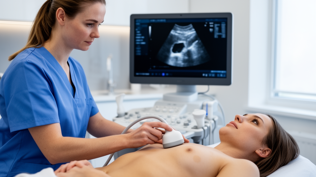
In adult cardiology departments, transthoracic echocardiography serves as the primary non-invasive imaging modality for evaluating cardiac anatomy and guiding clinical decision-making. To ensure that structural measurements accurately reflect physiological status rather than individual physical stature, clinical guidelines recommend indexing cardiac chamber dimensions and aortic diameters to patient height or body surface area. A prospective clinical service evaluation conducted across two National Health Service hospital sites in the North of England has demonstrated that while patients’ self-reported heights and weights differ statistically from objective measurements, these discrepancies lead to minimal clinically important alterations in echocardiographic classifications. The investigation showed that although patients tend to systematically overestimate their height and underestimate their weight, the overall correlation between reported and measured metrics remains exceptionally high. Consequently, calculated body surface area values derived from patient recall produce indexed cardiac dimensions that rarely cross diagnostic normality thresholds during routine clinical out-patient assessments.
The accurate quantification of cardiac chamber size represents a vital foundation in echocardiography, directly informing clinical management, risk stratification, and timely surgical interventions. Practice guidelines published by the British Society of Echocardiography, alongside joint recommendations from the American Society of Echocardiography and the European Association of Cardiovascular Imaging, emphasize comparing measured chamber dimensions against established reference ranges. Because anatomical chamber dimensions naturally vary according to subject-specific biological characteristics including sex, age, ethnicity, physical fitness, height, and overall body weight, clinical practice routinely indexes measurements to adjust for body size. In standard clinical workflows, body surface area is routinely derived from anthropometric values supplied directly by the patient rather than obtained via objective measurement. Despite this widespread operational reliance on patient memory, the actual accuracy of self-reported height and weight among outpatients attending echocardiography departments had remained unquantified, creating persistent clinical uncertainty regarding whether self-reported values might introduce misclassification into diagnostic reports.
To evaluate this diagnostic question, clinical researchers conducted a prospective service evaluation within the South Tees NHS Foundation Trust. The investigation was formally pre-registered, approved, and executed under an established service evaluation framework following consultations with the trust Research and Development department and guidance from the Health Research Authority decision tool. Consecutive adult outpatients attending for clinically indicated transthoracic echocardiography between March 22 and July 19, 2024, were enrolled after providing informed verbal consent. The evaluation encompassed patients attending dedicated cardiorespiratory departments at The James Cook University Hospital, a tertiary facility, and the Friarage Hospital, a secondary care centre. To preserve clinical flow and study consistency, patients attending community diagnostic centres or outpatient cardiology clinics were excluded for staffing reasons. In addition, individuals attending during fourteen days of clinical support worker industrial action were excluded from participation due to operational service constraints during those specific work stoppages.
A total of 698 adult patients participated in the evaluation across the two hospital sites, including 414 individuals recruited at The James Cook University Hospital and 284 patients at the Friarage Hospital. The overall cohort exhibited a representative demographic profile, containing 326 female participants, representing 46.7 percent, and 372 male participants, accounting for 53.3 percent, with a mean cohort age of 65.7 years and a standard deviation of 16.8 years. The study revealed marked feasibility differences between patient recall and physical measurement. While only 18 individuals, representing 2.6 percent of the total cohort, could not have their height or weight measured due to severe physical frailty or confinement to wheelchairs, 146 participants, representing 20.9 percent, were completely unable to provide a self-reported height, weight, or both prior to their examination. This demonstrated that measuring physical parameters is clinically feasible in almost all attending outpatients.
The proportion of participants unable to self-report height or weight differed substantially between the two hospital sites, illustrating how physical clinical environments influence patient communication. At The James Cook University Hospital, only 38 participants, or 9.0 percent of the local cohort, were unable to self-report their measurements, whereas at the Friarage Hospital, 108 individuals, representing 38.1 percent of patients, could not provide recall values. The study authors identified differences in physical data collection settings as the primary explanation for this disparity. At James Cook University Hospital, self-reported metrics were obtained inside private clinical rooms, whereas the Friarage Hospital lacked an equivalent private room, requiring measurements to be collected in a quiet corridor away from other individuals. Although staff maintained patient confidentiality, participants in semi-public corridor spaces may have experienced unease discussing body weight, opting instead to have healthcare professionals measure and record their parameters without verbal disclosure.
During data collection, participants first reported their height and weight in either metric or imperial units, after which staff measured their height and weight using a calibrated Marsden M-100 column weighing scale. Participants were weighed wearing light clothing, reflecting routine clinical practice in National Health Service outpatient departments. Body surface area was calculated using the Du Bois formula, multiplying 0.007184 by height raised to the 0.725 power and weight raised to the 0.425 power, yielding separate self-reported and measured surface area values for each individual. Transthoracic echocardiograms were performed and interpreted either directly by or under the immediate supervision of British Society of Echocardiography accredited level II adult echocardiographers using GE Vivid E95, GE Vivid S70, Philips Epiq, or Philips Affinity ultrasound machines. Indexed parameters were extracted retrospectively from authorized clinical reports stored within Philips IntelliSpace Cardiovascular reporting software. In accordance with British Society of Echocardiography guidelines, Sinuses of Valsalva and proximal ascending aorta diameters were indexed to height, while ventricular and atrial dimensions were indexed to body surface area.
Statistical comparisons revealed exceptionally strong correlations between self-reported and measured anthropometric values, with Pearson correlation coefficients exceeding 0.90 across all evaluations. Specifically, Pearson correlations reached 0.94 for height, 0.95 for weight, and 0.97 for calculated body surface area, with all comparisons reaching high statistical significance at p values below 0.001. Paired t-tests demonstrated systematic reporting biases across the cohort: mean self-reported height was 1.1 centimetres higher than measured height, averaging 169.5 centimetres compared to 168.4 centimetres, while mean self-reported weight was 1.6 kilograms lower than measured weight, averaging 80.8 kilograms compared to 82.4 kilograms. Because height overestimation and weight underestimation exert opposing mathematical influences within the Du Bois formula, the resulting mean body surface area discrepancy was restricted to 0.01 square metres, averaging 1.91 square metres for self-reported data versus 1.92 square metres for measured data, with a paired t-test p value of 0.008. These observed differences closely align with published literature where self-reporting discrepancies typically range from 0.5 to 1.3 centimetres for height and 1.1 to 1.5 kilograms for weight.
Indexing echocardiographic parameters against self-reported values generated minor but statistically significant differences across four specific anatomical structures. Indexed left ventricular end-diastolic volume averaged 49.3 millilitres per square metre using self-reported body surface area compared to 50.1 millilitres per square metre using measured values, representing a statistically significant difference with a small Cohen’s d effect size of 0.25. Indexed left ventricular end-systolic volume showed a similar slight difference of 21.3 versus 21.6 millilitres per square metre with an effect size of 0.19. Height-indexed Sinuses of Valsalva diameter averaged 18.6 millimetres per metre using self-reported height compared to 19.0 millimetres per metre using measured height with an effect size of negative 0.29, while proximal ascending aorta diameter averaged 18.4 versus 18.6 millimetres per metre with an effect size of negative 0.23. Conversely, left atrial volume, left ventricular mass, right atrial area, and right ventricular end-diastolic area demonstrated no statistically significant differences between self-reported and measured indexing approaches, confirming that the physiological impact of self-reporting remains narrow across routine chamber assessments.
Evaluation of diagnostic categorization under British Society of Echocardiography reference ranges showed that indexed parameter classifications remained unchanged in 95.9 to 100 percent of cases across all measured structures. No participant experienced a classification change spanning more than one diagnostic category, with shifts limited entirely to adjacent classifications. Proximal ascending aorta diameter proved most susceptible to reclassification, with 4.1 percent of patients crossing the diagnostic normality threshold, representing 18 out of 441 evaluated individuals. Specifically, 2.7 percent of cases shifted from abnormal to normal when using self-reported height, while 1.4 percent shifted from normal to abnormal. This directional asymmetry stems directly from the height-only indexing convention for aortic diameters, where over-reporting height increases the denominator and artificially reduces the indexed diameter. Other parameters demonstrated even higher diagnostic stability, with right atrial area remaining completely unchanged across all 467 evaluated patients, and left ventricular volumes showing zero cases crossing the normality threshold, demonstrating that routine diagnostic interpretation is largely preserved when self-reported metrics are utilized.
Subgroup analyses revealed that participants aged 60 to 80 years showed the greatest tendency to overestimate height, likely reflecting recall of peak lifetime stature rather than current standing height reduced by normal age-related musculoskeletal changes. While routine discrepancies were small, the study identified rare extreme outliers, including individual weight errors exceeding 30, 40, and 60 kilograms, and height discrepancies surpassing 10, 20, and 30 centimetres. The authors also noted ongoing clinical debates regarding body surface area indexing in obesity, where formula inflation can mask left atrial enlargement and diastolic dysfunction. Indications for surgical intervention on the ascending aorta depend upon absolute dimensions rather than indexed values, mirroring British Society of Echocardiography advice to prioritize absolute left ventricular dimensions. Ultimately, the investigators concluded that while objective measurement remains the guideline-recommended gold standard—achievable in 97.4 percent of patients—self-reported height and weight provide an accurate and dependable clinical alternative for most outpatients attending transthoracic echocardiography, provided that practitioners remain vigilant in identifying rare individuals who significantly misreport physical dimensions.
Subject of Research: Accuracy of self-reported height and weight in adult transthoracic echocardiography
Article Title: Discrepancies and potential impacts of self-reported versus measured height and weight on adult transthoracic echocardiography findings
Article References: Benson, C., Austin, D., Graham, R., & Wilkinson, C. (2025). Discrepancies and potential impacts of self-reported versus measured height and weight on adult transthoracic echocardiography findings. Echo Research & Practice, 12(1), Article 31. https://doi.org/10.1186/s44156-025-00095-y
Image Credits: AI Generated
DOI: 10.1186/s44156-025-00095-y
Keywords: transthoracic echocardiography, body surface area, cardiac imaging, clinical cardiology, anthropometric measurements, echocardiographic chamber quantification, left ventricular volume, aortic diameter, Discrepancies, potential, impacts, self-reported
News Source: Ophelia Keating. (November 10, 2026). Self-Reported Patient Measurements Accurately Guide Most Routine Echocardiogram Classifications. Scienmag.