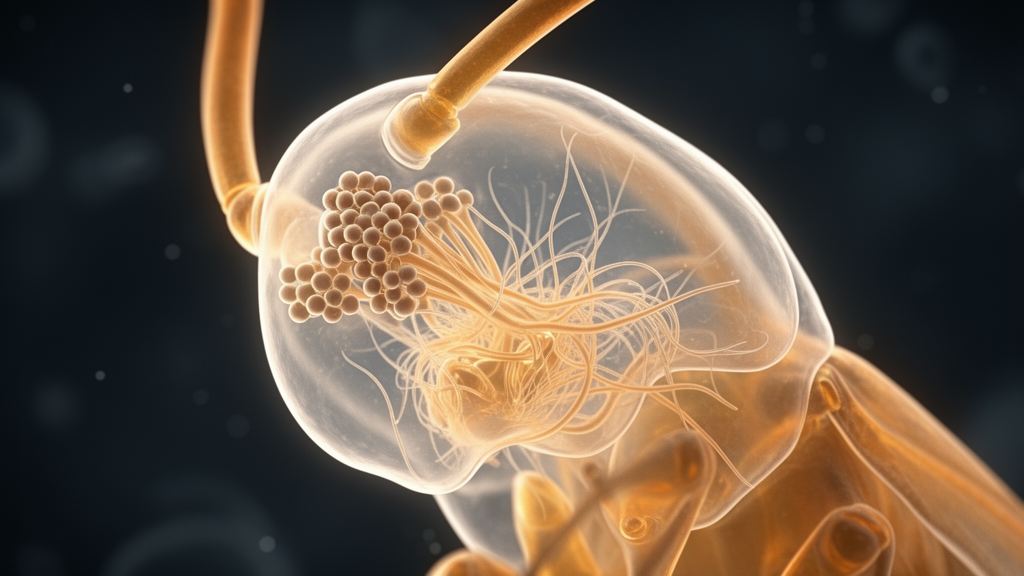
Researchers have detailed the early embryonic development of Johnston’s organ in the desert locust, Schistocerca gregaria, identifying the specific cellular precursors that build this auditory structure. Published in Development Genes and Evolution, the study by George Boyan and Erica Ehrhardt provides the first modern developmental account of how this sensory organ forms in a hemimetabolous insect. Because locusts lack a pupal stage, their sensory organs must be fully functional upon hatching, necessitating that Johnston’s organ develops entirely during embryogenesis. The team used immunolabeling and confocal microscopy to track the cellular origins of the organ within the antenna’s pedicellar segment.
The study focused on the pedicel, the short intermediary segment of the locust antenna that houses Johnston’s organ. In adult locusts, this organ consists of symmetrical dorsal and ventral cell clusters, each containing six to seven scolopales that insert into a membrane at the pedicellar-flagellar junction. The researchers aimed to establish the epithelial domain of origin for these structures and analyze the growth of the neuronal lineages they generate. By screening 235 preparations, the team determined that the major developmental steps building the organ occur prior to mid-embryogenesis, with the cellular organization at this stage already reflecting the adult structure.
To identify the specific region of the antenna where the organ develops, the researchers employed the epithelial cell marker Lachesin. This GPI-linked cell surface molecule is expressed initially on all differentiating epithelial cells but is retained later only by cells involved in neurogenesis. At mid-embryogenesis, Lachesin expression in the scape and pedicel matched the segmentation revealed postembryonically by epifluorescence illumination. Even earlier, at 32% of embryogenesis, a distinct stripe of Lachesin-positive cells marked the epithelial domain of the pedicel, allowing the team to unambiguously identify the region before the organ was fully formed.
The team utilized a triple-labeling protocol involving Lachesin, the mitosis marker phosphohistone-3, and neuron-specific horseradish peroxidase. This approach allowed them to visualize mitotically active precursors associated with the epithelial domains in the flagellum and pedicel. At 34% of embryogenesis, Lachesin-positive cell clusters of the future Johnston’s organ were visible in the pedicel, while no differentiated neurons were present in the pedicel itself. This method provided a clear molecular basis for tracking the cellular development of the organ from its earliest origins within the identifiable pedicellar region.
At approximately 36% of embryogenesis, a single large Lachesin-positive and phosphohistone-3-positive mitotically active cell was identified in the midline region of the pedicellar domain. This sense-organ precursor, or SOP, was itself horseradish peroxidase-negative but had generated a small lineage of four Lachesin-positive progeny. Two of these progeny were horseradish peroxidase-positive, representing the initial neurons of the cell cluster. As development progressed to 42%, three phosphohistone-3-positive mitotically active cells were observed generating lineages in the ventral domain. By 48% of embryogenesis, six progenitors were distributed throughout the ventral domain, matching the complement of cell clusters found in the mature organ.
The study revealed that the locations of these sense-organ precursors are remarkably conserved across different preparations and ages. This consistency suggests that the epithelium possesses an underlying topographic coordinate system that determines the cellular organization of Johnston’s organ. By mid-embryogenesis, twelve lineages are arranged circumferentially in the pedicel, mirroring the adult structure. The spatial distribution of clusters within a domain changes in a binary fashion with age, and cluster numbers are remarkably constant across all preparations at each given age, with no variance observed at specific developmental stages.
Each sense-organ precursor is associated with a smaller mitotically active cell from which the neuronal complement of each clone may derive. The researchers established that there is only a single large SOP associated with each progressively expanding lineage, indicating that the progeny represent a single clone. Neuron numbers within a clone increase in discrete steps with age, stabilizing at five cells per clone by 40% of embryogenesis. These neuronal progenies are consolidated into tightly bound cartridges, a structure reminiscent of retinal photoreceptors in flies and other sensory units in insects.
A long scolopale extends apically from each cartridge to an insertion point in the epithelium, while bundled axons project basally toward the brain. At 55% of embryogenesis, scolopales from all six ventral and six dorsal cell clusters extend in parallel for over 50 micrometers toward the flagellum. The neurons have generated extensive dendritic processes projecting apically and axons projecting basally to the scape, where they fasciculate with the antennal nerve. This structural organization at mid-embryogenesis strongly resembles that of the adult, suggesting that subsequent development involves maturation rather than the generation of additional lineages.
The findings indicate that the desert locust’s Johnston’s organ possesses a type I sensory organization common to other internal chordotonal organs for stretch or vibration. Comparative data reveal a similar circumferential organization of cell clusters in the embryonic locust and the phylogenetically distant adult ant, consistent with a conserved structural requirement for detecting environmental signals. The study highlights that the mechanisms regulating the developmental program of Johnston’s organ in the locust may be homologous to those identified in Drosophila, involving factors such as homothorax, cut, and atonal, though this remains to be fully clarified.
Despite the detailed mapping of neuronal lineages, the identity of additional non-neuronal cells associated with each cartridge remains unclear. Double-labeling with the sensory cell marker Lazarillo revealed extrinsic cells that are exclusively horseradish peroxidase-negative and Lazarillo-positive. These cells could represent attachment or ligament cells found in other insects, but their specific identity in the locust requires further investigation. The study provides a foundational framework for understanding how topographic coordinates and cellular lineages combine to form a complex auditory organ during embryonic development.
Detailed staging methods and antibody staining specifics formed the experimental foundation of the study. The researchers sourced eggs from a crowded colony of desert locusts and staged embryos to the nearest one percent of developmental time, where five percent corresponds to twenty-four hours of development. To visualize the cellular machinery, the authors combined distinct immunological probes targeting specific molecular markers. Neuron-specific anti-horseradish peroxidase recognized insect neural epitopes, whereas an anti-Lachesin antibody targeted a cell surface molecule of the immunoglobulin superfamily that marks differentiating epithelium and persists in neurogenic precursors. Mitotically dividing cells were pinpointed using an antibody against phospho-histone H3, which binds the phosphorylated amine terminal of Histone 3 during chromosome condensation, exhibiting its strongest signal during metaphase. To track non-neuronal elements, the team incorporated an antibody against Lazarillo, a surface lipocalin expressed by sensory cells. Visual detection was achieved through secondary antibodies conjugated to fluorophores including Alexa 488, Cy3, and Cy5, alongside peroxidase conjugation counterstained with diaminobenzidine under Nomarski differential interference contrast optics.
Quantitative evaluation of the lineages demonstrated strict developmental pacing across dozens of individual embryos. The investigators tracked cluster counts and neuron counts across multiple embryonic stages, analyzing seven preparations at 36 percent of embryogenesis, four at 38 percent, eleven at 39 percent, eighteen at 40 percent, six at 41 percent, six at 45 percent, four at 48 percent, four at 50 percent, and four at 55 percent. Across all eighteen preparations examined at the 40 percent mark, the number of identifiable clusters exhibited zero variance. Transverse optical reconstructions documented how the spatial distribution expanded symmetrically in three dimensions. At 41 percent of embryogenesis, optical cross-sections through the pedicel revealed three ventral neuronal clusters paired with an identical mirror-symmetrical group of three dorsal clusters. By 55 percent of embryogenesis, both the ventral and dorsal domains had doubled their complement to six clusters each, completing a circumferential ring of twelve cartridges that matches the configuration found in mature locust antennae.
The progression of individual cartridges followed an equally orderly schedule before stabilizing prior to mid-embryogenesis. Single-cell reconstructions showed that the neuronal yield per clone expanded sequentially from two differentiated neurons at 36 percent of development to three at 38 percent, four at 40 percent, and reached a plateau of five cells at 41 percent. No statistically significant difference in neuron counts was detected between the ventral and dorsal clusters at any sampled stage. Alongside the developing cartridges of Johnston’s organ, the pedicellar epithelium hosted other emerging sensory components, including chordotonal organs and laterally situated campaniform sensilla. Axons from flagellar pioneer neurons coursed through the pedicel in parallel ventral and dorsal tracts toward the brain, while the newly differentiated sensory neurons of Johnston’s organ projected their own axonal bundles into scapal nerves and extended apical scolopales toward cap cell attachment points.
These observations underscore significant developmental divergences between hemimetabolous insects and holometabolous model organisms such as Drosophila. In fruit flies, Johnston’s organ is assembled during the larval and pupal stages of metamorphosis, achieving functional auditory capacity only in the adult. In contrast, the hemimetabolous locust constructs its entire auditory apparatus during embryonic life so that sensory perception is operative immediately upon emergence from the egg. The authors noted that identifying the exact role of the smaller proliferative cell associated with each sense-organ precursor remains an important challenge. In Drosophila, secondary divisions follow an established pathway involving secondary precursor cells, but confirming whether a similar intermediate precursor lineage operates in locusts will require future lineage tracing using pulsed nucleotide analogs like bromodeoxyuridine or intracellular dye fills. Furthermore, resolving the identity of the non-neuronal Lazarillo-positive cells within each cartridge may require specialized markers such as alpha-tubulin for attachment cells or fluorescent phalloidin for scolopales.
Subject of Research: Embryonic development of Johnston's organ in the desert locust antenna
Article Title: Early embryonic development of Johnston’s organ in the antenna of the desert locust Schistocerca gregaria
Article References: Boyan, G., & Ehrhardt, E. (2022). Early embryonic development of Johnston’s organ in the antenna of the desert locust Schistocerca gregaria. Development Genes and Evolution, 232(5-6), 103-113. https://doi.org/10.1007/s00427-022-00695-2
Image Credits: AI Generated
DOI: 10.1007/s00427-022-00695-2
Keywords: Johnston's organ, desert locust, embryonic development, antenna, sensory organ, cell lineage, immunolabeling, insect biology, neurogenesis, topographic organization, Early, embryonic
News Source: Drew Townsend. (November 10, 2026). Locust Antenna Hearing Organ Forms During Embryonic Development. Scienmag.