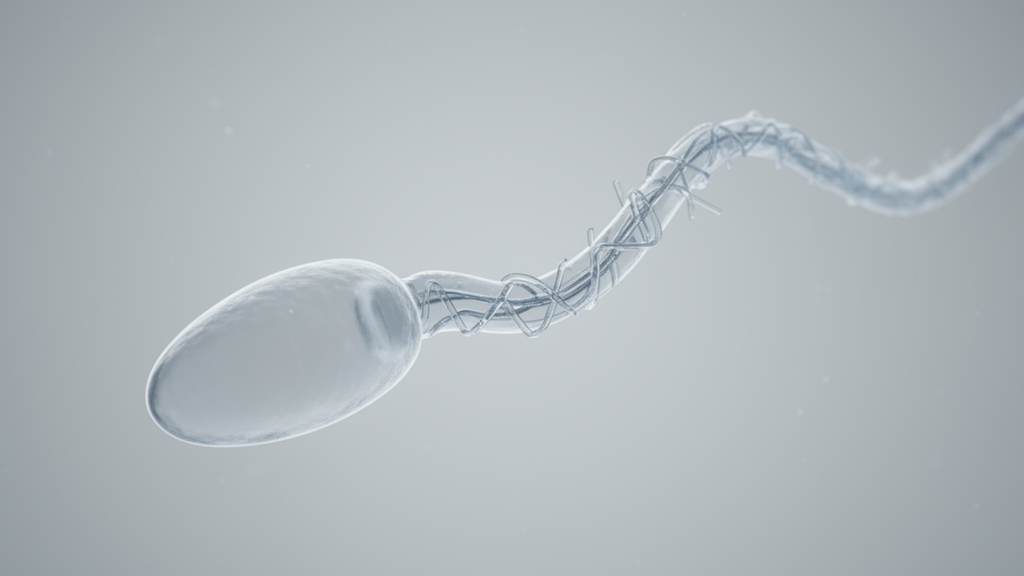
Researchers identified a novel homozygous splice-site variant in the CFAP65 gene that is associated with oligozoospermia and complete asthenozoospermia in a consanguineous family. The study, published in BMC Medical Genomics, investigates how this specific genetic alteration affects sperm function through aberrant splicing mechanisms. The proband, a 36-year-old man with a ten-year history of primary infertility, presented with severe sperm motility defects. Semen analysis revealed a sperm concentration of 8.1 × 10⁶/mL, which is significantly below normal ranges, indicating oligozoospermia. Furthermore, the analysis showed that total motility and progressive motility were both 0%, while 100% of the spermatozoa were immotile. These clinical findings are consistent with complete asthenozoospermia, a condition characterized by the total lack of sperm movement.
The investigation utilized whole-exome sequencing to identify candidate variants in the proband and his parents. This genomic approach allowed the team to filter variants based on clinical phenotype, disease databases, and population allele frequency data. The analysis focused on identifying variants that could explain the severe motility defects observed in the patient. The CFAP65 gene encodes a testis-enriched protein essential for maintaining sperm flagellar integrity. It is proposed to participate in the assembly and stabilization of the axonemal dynein regulatory complex, a structure critical for regulating flagellar beating. Biallelic pathogenic variants in CFAP65 have previously been shown to disrupt this architecture, leading to multiple morphological abnormalities of the sperm flagella and complete sperm immotility.
Whole-exome sequencing identified a novel homozygous canonical splice-site variant in CFAP65, designated as NM_194302.4:c.645 + 1G > A. This variant affects the invariant +1 nucleotide at the exon 6-intron 6 junction, a region critical for proper pre-mRNA splicing. Sanger sequencing confirmed that the proband was homozygous for this variant, while both parents were heterozygous carriers. This inheritance pattern is consistent with an autosomal recessive disorder. The variant was not recorded in ClinVar or reported in the literature at the time of analysis, highlighting its novelty. According to the American College of Medical Genetics and Genomics guidelines, the variant was classified as pathogenic based on criteria including its canonical splice-site location, rarity in population databases, and homozygous occurrence in an affected individual.
To assess the familial relatedness and support the autosomal recessive inheritance pattern, the researchers performed kinship and runs of homozygosity analyses. Genome-wide single-nucleotide variant data were used to calculate the genomic inbreeding coefficient, known as FROH. The analysis showed that the proband had an increased autosomal homozygosity, with an FROH value of 7.88%. This value corresponds to a total autosomal runs of homozygosity length of approximately 227 megabases. This level of homozygosity is compatible with the expected autozygous proportion for offspring of a first-cousin union, which is approximately 6.25%. The family history confirmed that the proband’s parents were first cousins, providing clinical context for the genomic findings. These results suggest that parental consanguinity increased the likelihood of the proband inheriting two copies of the pathogenic CFAP65 variant.
Functional assessment of the variant’s impact on splicing was conducted using both in silico prediction and in vitro experimental validation. The RNA Splicer tool, which integrates deep-learning-based models, predicted two aberrant splice patterns for the c.645 + 1G > A variant. The first pattern involved the creation of a novel donor site at exon 6, leading to a 5-bp insertion and a subsequent frameshift. The second pattern predicted complete skipping of exon 6, resulting in a 103-bp deletion from the transcript. To experimentally validate these predictions, the researchers performed a minigene splicing assay in HEK-293T cells. This assay involved constructing minigene plasmids containing genomic sequences spanning CFAP65 exons 5 through 7, with either the wild-type or mutant allele.
The minigene assay provided definitive evidence for the splicing consequence of the variant. Reverse transcription-polymerase chain reaction analysis showed that cells transfected with the wild-type construct produced a 529-bp amplicon, representing the normal transcript. In contrast, cells transfected with the mutant construct generated a 426-bp amplicon, indicating a reduction in transcript size. Sanger sequencing of the PCR products confirmed that the c.645 + 1G > A variant caused complete skipping of exon 6. This skipping resulted in a 103-bp deletion in the transcript, leading to a predicted frameshifted truncated protein designated as p.(Pro182ArgfsTer29). The frameshift occurs at residue 182, which is highly conserved across species, suggesting that this region is functionally important for the protein’s structure and activity.
The predicted truncated protein is expected to undergo nonsense-mediated decay, a cellular mechanism that degrades mRNA transcripts containing premature stop codons. This splice-mediated loss-of-function mechanism is consistent with previous reports showing that biallelic loss-of-function variants in CFAP65 are associated with severe asthenozoospermia and sperm flagellar abnormalities. The study expands the known mutational spectrum of CFAP65 by providing functional evidence for a canonical splice-site variant. While previous studies have identified various CFAP65 variants, transcript-level functional validation for splice-region variants had been limited. This study bridges that gap by demonstrating the specific splicing defect caused by the c.645 + 1G > A variant.
The clinical presentation of the proband included not only motility defects but also abnormal sperm morphology. The total abnormal morphology rate was 88.0%, with a head defect rate of 85.2%. Despite the complete absence of motility, sperm vitality remained at 51.7%, indicating that a substantial proportion of spermatozoa were viable but immotile. This distinction is clinically relevant for assisted reproduction, where assessment of sperm viability is important when considering immotile sperm for use. However, detailed sperm morphology classification and transmission electron microscopy analysis were not performed in this study. Therefore, direct comparison with the specific flagellar subtypes and ultrastructural features reported in prior CFAP65 cases is limited. The researchers noted that whether the patient fully meets the criteria for multiple morphological abnormalities of the sperm flagella cannot be determined without comprehensive ultrastructural assessment.
The study highlights the value of integrating genomic and pedigree information in the diagnosis of male infertility, particularly in consanguineous families. The combination of whole-exome sequencing and runs of homozygosity analysis proved to be an effective strategy for prioritizing candidate genes and variants. The kinship analysis confirmed the parent-child relationships and indicated increased genetic similarity between the parents, supporting the documented consanguinity. This integrated approach allows for a more accurate assessment of the genetic basis of infertility in affected families. The findings also underscore the importance of genetic risk assessment and counseling for families with a history of consanguinity and reproductive issues.
Methodologically, the study demonstrates the utility of combining in silico prediction with functional validation for splice-site variants. The RNA Splicer tool provided initial predictions of aberrant splicing patterns, which were then confirmed through the minigene assay. This integrated approach ensures that the functional consequences of genetic variants are accurately characterized. However, the researchers acknowledged limitations in the study design. Because CFAP65 is highly and preferentially expressed in the testis, reverse transcription-polymerase chain reaction validation using patient-derived peripheral blood RNA could not be performed. The minigene assay was conducted in HEK-293T cells, a cell line that may not fully recapitulate tissue-specific splicing regulation in testicular germ cells. Future studies using appropriate reproductive samples or germ-cell-related models would be important to further clarify the downstream molecular consequences of CFAP65 deficiency.
The findings of this study contribute to the understanding of the genetic etiology of male infertility with sperm motility defects. By identifying a novel homozygous CFAP65 splice-site variant and demonstrating its functional impact, the researchers provide new insights into the pathogenic mechanisms underlying asthenozoospermia. The study emphasizes the need for comprehensive genetic screening and functional splicing assessment in clinical settings.
Subject of Research: Genetic basis of male infertility involving CFAP65 splice-site variants and sperm motility defects
Article Title: Identification and splicing assessment of a novel homozygous CFAP65 splice-site variant associated with oligozoospermia and complete asthenozoospermia
Article References: Teng, C., Tang, X., Li, L., Ma, Y., Zhao, Y., Chen, Q., Shi, H., & Zhang, J. (2026). Identification and splicing assessment of a novel homozygous CFAP65 splice-site variant associated with oligozoospermia and complete asthenozoospermia. BMC Medical Genomics, 19(1), Article 155. https://doi.org/10.1186/s12920-026-02466-8
Image Credits: AI Generated
DOI: 10.1186/s12920-026-02466-8
Keywords: CFAP65, male infertility, asthenozoospermia, oligozoospermia, splice-site variant, whole-exome sequencing, minigene assay, consanguinity, sperm motility, genetic diagnosis, Identification, splicing
News Source: Ophelia Keating. (November 10, 2026). Novel CFAP65 Splice Variant Linked to Complete Sperm Immotility. Scienmag.