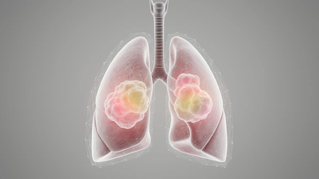
Researchers have developed a deep learning framework that computationally visualizes elastic fiber architecture from standard hematoxylin and eosin (H&E) images, aiming to improve the objectivity of histological grading in lung adenocarcinoma. The study, published in Diagnostic Pathology, addresses a significant limitation in current pathology practices: the high interobserver variability associated with distinguishing noninvasive lepidic growth from invasive patterns. This distinction is critical for accurate grading but often difficult to achieve on routine H&E slides because the alveolar elastic fiber framework, which serves as a key structural marker, is frequently insufficiently visualized without specialized staining.
The core innovation of this work is the creation of a “virtual” imaging pipeline that eliminates the need for additional staining or fluorescence microscopy. The team developed a dual-stream EfficientNet-B0 classifier trained on spatially coregistered pairs of standard H&E images and eosin-based elastin fluorescence (EBEF) images. To enable clinical deployment in standard pathology workflows, they employed a Pix2Pix-HD generative adversarial network to synthesize high-fidelity virtual EBEF images directly from H&E inputs. This approach allows the automated pipeline to classify histological patterns, quantify the proportions of different growth patterns, and assign tumor grades using only the routine slides already available in clinical settings.
The performance of the system was evaluated on independent multicenter cohorts to ensure robustness. The benchmark dual-stream model, which used real H&E and EBEF images, achieved a tile-level classification accuracy of 89.3%. When the automated grading pipeline utilized the synthesized virtual EBEF images, it achieved an accuracy of 94.2% on an external validation cohort. This performance significantly outperformed a model relying solely on H&E images, which recorded an accuracy of 79.6%.
Ablation studies were conducted to determine which components of the pipeline contributed most to the performance gains. The analysis confirmed that the improvements depended on three key factors: the use of informative multimodal input, accurate spatial registration between the H&E and EBEF images, and attentive feature fusion within the neural network architecture.
The study also explored the prognostic relevance of the AI-assigned grades using a curated cohort from The Cancer Genome Atlas (TCGA) lung adenocarcinoma dataset. The results demonstrated a consistent trend toward an association between the assigned grades and overall survival. Specifically, significant stratification was observed when low-grade tumors were combined, with a log-rank p-value of 0.048.
By enabling objective grading from a single routine H&E slide, the approach aims to reduce the subjectivity inherent in manual histological assessment. The ability to enhance the visualization of elastic fiber-related architecture computationally means that pathologists can potentially achieve greater consistency in grading without altering their existing procedures or requiring additional laboratory resources.
Subject of Research: Deep learning framework for virtual elastic fiber imaging in lung adenocarcinoma grading
Article Title: Deep learning-powered virtual eosin-based elastic fiber framework imaging improves objective grading and prognostic stratification of lung adenocarcinoma
Article References: Wang, C.-L., Lei, S.-P., Lu, Y., Zou, L.-F., Xiong, J.-B., Li, J.-W., Ouyang, X., He, L., Luo, Y.-Y., Zhang, L., Cao, X.-J., & Yu, S.-S. (2026). Deep learning-powered virtual eosin-based elastic fiber framework imaging improves objective grading and prognostic stratification of lung adenocarcinoma. Diagnostic Pathology. https://doi.org/10.1186/s13000-026-01837-0
Image Credits: AI Generated
DOI: 10.1186/s13000-026-01837-0
Keywords: lung adenocarcinoma, deep learning, histological grading, virtual imaging, pathology, prognostic stratification, H&E staining, elastic fiber, artificial intelligence, medical imaging, Deep, learning-powered
News Source: Ophelia Keating. (November 10, 2026). AI Virtual Imaging Improves Lung Cancer Grading. Scienmag.