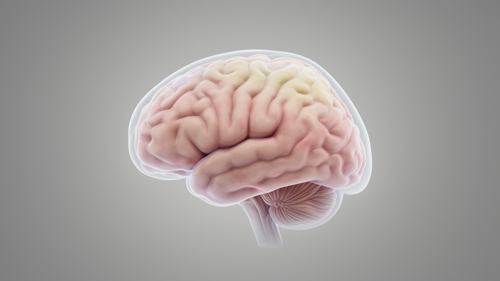
A study published in BMC Medical Imaging reported cortical and subcortical morphometric differences in adolescents with first-episode, medication-naïve early-onset depression. The study, published in November 2026, utilized surface-based morphometry to analyze structural MRI data from 161 adolescents with early-onset depression and 161 healthy controls. The primary objective was to investigate specific morphometric alterations in the cortex and subcortical regions, providing a detailed map of structural variations associated with the condition in this specific demographic.
The analytical approach relied on FreeSurfer software to process the structural MRI data, generating vertex-wise maps that were smoothed with a 10-mm full width at half maximum kernel. To ensure statistical rigor, the researchers adjusted for age, sex, intelligence quotient, and estimated total intracranial volume. Cluster-wise corrections were applied using 1000 permutations to account for multiple comparisons at the vertex level. Additionally, parcel-level group differences were assessed using general linear models, with Benjamini–Hochberg false discovery rate correction applied within each morphometric domain to control for false positives.
The vertex-wise analyses revealed a complex pattern of cortical changes, characterized by widespread cortical thickening, broadly reduced cortical surface area, and mixed changes in cortical volume. These findings were mirrored in the parcel-level analyses, which also indicated predominantly increased cortical thickness and reduced cortical surface area. The subcortical structures displayed distinct volumetric differences, including lower bilateral pallidal and right hippocampal volumes. Conversely, the study found higher volumes in the bilateral accumbens, right thalamic, and right amygdala regions.
In an exploratory analysis, the team examined the relationship between specific morphometric measures and symptom severity within the depression group. Partial correlations were calculated with Hamilton Rating Scale for Depression 17-item scores, controlling for the same covariates used in the primary analyses. The results showed that left entorhinal thickness was inversely associated with symptom scores, with a partial correlation coefficient of −0.298 and a q-value of 0.0255 under the exploratory correction. However, this association did not survive a more stringent sensitivity correction that combined both symptom scales.
The study’s cross-sectional design imposes significant limitations on the interpretation of these findings. While the sample demonstrated clear group-level morphometric differences, the data cannot establish whether these structural changes represent altered developmental trajectories or are closely proximal to the onset of the illness. The authors explicitly noted that the study does not support claims regarding individual diagnostic utility or treatment selection.
Subject of Research: Cortical and subcortical morphometric differences in adolescents with early-onset depression
Article Title: Neurodevelopmental cortical morphometric alterations in medication-naïve adolescents with first-episode early-onset depression: a surface-based morphometry study
Article References: Subi, Y., Kukun, H., Tuxunjiang, P., Aihemaiti, A., Xu, R., Li, S., Jiang, J., Yan, P., Sheng, W., & Wang, Y. (2026). Neurodevelopmental cortical morphometric alterations in medication-naïve adolescents with first-episode early-onset depression: a surface-based morphometry study. BMC Medical Imaging. https://doi.org/10.1186/s12880-026-03025-4
Image Credits: AI Generated
DOI: 10.1186/s12880-026-03025-4
Keywords: adolescent depression, neuroimaging, cortical thickness, surface area, subcortical volumes, FreeSurfer, structural MRI, early-onset depression, morphometry, medication-naïve, Neurodevelopmental, cortical
News Source: Glenn Wilkins. (November 9, 2026). Brain Scans Reveal Cortical Differences in Adolescent Depression. Scienmag.