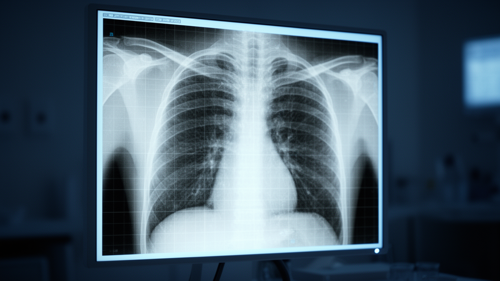
A deep learning framework utilizing frozen visual features extracted from standard chest radiographs can identify moderate-to-severe anemia in adult patients without requiring dedicated model fine-tuning, according to a retrospective hospital cohort study conducted in Taiwan. The research team evaluated whether noninvasive imaging could flag clinically meaningful hemoglobin deficits by capturing subtle secondary cardiovascular adaptations and radiodensity shifts across thoracic structures. Using a vast hospital archive containing hundreds of thousands of imaging records, the investigators demonstrated that visual representations derived from routine chest X-rays can differentiate patients with hemoglobin levels of 10.0 grams per deciliter or lower from those with higher concentrations. Because hemoglobin evaluation traditionally mandates invasive venipuncture, laboratory infrastructure, and specialized personnel that may not be readily available in remote clinical environments or mobile health units, opportunistic evaluation of already-acquired radiographic examinations presents a practical pathway for automated risk stratification and early clinical notification during routine hospital care.
The study cohort was drawn from electronic health records and picture archiving systems at Tri-Service General Hospital in Taipei, Taiwan, covering consecutive adult encounters recorded between June 2016 and February 2022. Eligible participants were at least 20 years of age and had undergone at least one chest X-ray during an emergency department visit, outpatient clinic consultation, or inpatient ward admission. To simulate realistic real-world clinical presentations and avoid over-representing repeat imaging events, investigators selected exactly one chest radiograph per patient at a randomized time point. In total, 305,793 adult patients met the inclusion criteria. The dataset was partitioned into four non-overlapping subsets: a development cohort of 135,867 radiographs and an optimization tuning cohort of 54,405 radiographs used for model training and parameter adjustments, an internal validation cohort comprising 67,623 radiographs from hospital branches outside Jingjhou, and an independent external validation cohort of 47,898 radiographs collected exclusively from the hospital’s Jingjhou branch to test generalizability across an independent medical facility.
To convert complex two-dimensional radiographic images into computable numerical features, the investigators implemented an image-text contrastive architecture based on the Pretraining Approach for Small-sample Training Employing Radiographs framework. High-resolution digital imaging files stored in standard DICOM format were resized to 256 by 256 pixels and processed through a pretrained Vision Transformer encoder, specifically the ViT-B/32 architecture. The model utilized fixed pretrained weights without additional fine-tuning or end-to-end retraining on the hospital dataset. Passing each image through the Vision Transformer produced a dense 512-dimensional vector embedding summarizing key anatomical and structural visual characteristics. These visual embeddings served directly as input variables for a downstream logistic regression classifier, termed the CXR-risk score, specifically calibrated to predict moderate-to-severe anemia, which was defined as a laboratory-confirmed hemoglobin concentration of 10.0 grams per deciliter or below measured within 24 hours of the radiographic exposure.
Across the independent testing sets, the standalone chest radiograph framework demonstrated consistent discriminative capacity. In the internal validation cohort of 67,623 patients, where the baseline anemia prevalence was 8.8 percent, the model achieved an area under the receiver operating characteristic curve of 0.845, accompanied by a sensitivity of 68.5 percent, a specificity of 84.7 percent, a positive predictive value of 30.0 percent, and a negative predictive value of 96.5 percent. In the external validation cohort of 47,898 patients from the Jingjhou branch, with a baseline anemia prevalence of 9.0 percent, the system attained an area under the curve of 0.852, a sensitivity of 71.5 percent, a specificity of 83.2 percent, a positive predictive value of 29.5 percent, and a negative predictive value of 96.7 percent. Global probability calibration assessments further confirmed model reliability across both validation cohorts, producing standard Brier scores of 0.06306 internally and 0.06326 externally, reflecting scaled Brier reductions in overall predictive uncertainty of 21.28 percent and 22.52 percent respectively.
The researchers also benchmarked the imaging framework against clinical prediction models and combined multimodal approaches trained with gradient-boosted decision trees using the XGBoost algorithm. A predictive model relying exclusively on demographic and clinical variables, including age, sex, and underlying diagnostic histories, achieved an area under the curve of 0.808 with a 95 percent confidence interval of 0.802 to 0.814 in the internal validation set, and 0.828 with a 95 percent confidence interval of 0.822 to 0.835 in the external cohort. In comparison, the standalone deep learning imaging score alone achieved superior discriminative accuracy over clinical features. Crucially, fusing the 512-dimensional radiographic embeddings with patient clinical data yielded the highest overall predictive accuracy, elevating the area under the curve to 0.878 with a 95 percent confidence interval of 0.873 to 0.882 internally, and to 0.881 with a 95 percent confidence interval of 0.876 to 0.887 in the external validation cohort, indicating that the visual representations capture independent physiopathological information not reflected by conventional electronic health record variables alone.
Subgroup analyses revealed notable variations in predictive accuracy across demographic segments, diagnostic hardware, projection views, and hospital departments. Predictive performance was higher when interpreting computed radiography images, yielding an area under the curve of 0.869 in internal testing and 0.867 externally, compared to digital radiography systems. Standard posteroanterior projections yielded higher discrimination than anteroposterior views in the internal dataset, reaching an area under the curve of 0.812, though this positional difference attenuated in external validation to an area under the curve of 0.816. Inpatient examinations yielded higher discrimination than outpatient or emergency department scans, achieving an area under the curve of 0.851 internally and 0.855 externally. By demographic group, the framework achieved its strongest performance among male patients, with an area under the curve of 0.909 internally and 0.910 externally, and individuals aged 55 to 64 years, reaching an area under the curve of 0.876 across both validation cohorts. Conversely, predictive accuracy dropped noticeably among younger female patients under 55 years of age.
The authors discussed the physiological mechanisms connecting severe systemic hemoglobin depletion with observable features on thoracic imaging. When hemoglobin concentrations remain above 10.0 grams per deciliter, non-hemodynamic cellular adaptations such as increased 2,3-diphosphoglycerate facilitate rightward shifts in oxygen dissociation to preserve peripheral oxygenation. However, once hemoglobin declines below the critical threshold of 10.0 grams per deciliter, significant compensatory cardiovascular alterations are triggered. Hypoxia-mediated vasodilation reduces systemic vascular resistance and cardiac afterload, while enhanced venous return augments ventricular preload. Through the Frank-Starling mechanism and sympathetic nervous system activation, stroke volume and inotropic contractility increase to maintain tissue perfusion. To examine which thoracic regions drove model predictions, the investigators generated exploratory feature visualization heatmaps. Prominent activation clustered over the cardiac silhouette, the pulmonary arteries, and the bilateral hilar regions, suggesting the network reacted to structural remodeling, blood pool redistribution, and vascular prominence induced by the hyperdynamic circulatory state.
Variable importance analysis conducted on integrated models indicated that underlying systemic disease histories and specific radiographic findings heavily influenced risk estimations. A documented history of chronic kidney disease emerged as the single most influential clinical predictor of anemia. In the study cohort, patients with chronic kidney disease exhibited an anemia prevalence of 37.4 percent, representing a 6.8-fold increase compared to individuals without kidney disease, among whom the prevalence was only 5.5 percent. Chronological age represented the second most important feature, exhibiting 64.4 percent relative importance compared to chronic kidney disease history. Image acquisition projection, distinguishing between posteroanterior and anteroposterior views, contributed 49.6 percent relative importance, followed by visible supportive medical devices such as nasogastric tubes at 38.3 percent relative importance and costophrenic angle blunting at 32.4 percent relative importance.
Because medical hardware and severe chronic organ damage can introduce confounding visual patterns, the investigators conducted extensive sensitivity evaluations to test whether the neural network relied on trivial artifacts or genuine physiological manifestations. In an exclusion experiment removing all radiographs containing visible foreign medical devices, including nasogastric tubes and indwelling Port-A catheters, model performance declined only modestly, shifting from an area under the curve of 0.845 to 0.813 in internal validation and from 0.852 to 0.817 in external testing. Furthermore, in a disease-free subgroup analysis that excluded all patients diagnosed with major cardiopulmonary conditions such as heart failure, coronary artery disease, chronic obstructive pulmonary disease, hypertension, and chronic kidney disease, the model retained an area under the curve of 0.81 and an external sensitivity of 0.75 across tens of thousands of examinations. These findings confirmed that the deep learning algorithm recognized radiographic signatures linked directly to hemoglobin deficits rather than merely detecting secondary devices or overt terminal cardiopulmonary failure.
To evaluate prospective clinical deployment, the researchers conducted decision curve analysis comparing model-guided blood testing against universal testing and zero-testing strategies. The deep learning framework delivered a superior net clinical benefit across threshold probabilities spanning 10 percent to 60 percent, demonstrating that triggering confirmatory blood tests based on radiographic risk scores could optimize diagnostic yields while averting unneeded laboratory procedures within that range. When operating under a high-risk operational cutoff, the framework achieved a specificity between 0.93 and 0.94 and raised the positive predictive value to 44 percent. Conversely, across unselected disease-free cohorts where comorbidity prevalence is low, the positive predictive value fell to between 0.12 and 0.16, meaning approximately 85 percent of positive model flags represented false positives, even though negative predictive values remained exceptionally high at 0.97 to 0.98. Consequently, the authors emphasized that the tool is fundamentally structured for opportunistic clinical screening and rapid rule-out assessments rather than definitive diagnostic replacement.
The authors outlined several methodological constraints inherent to the retrospective design and hospital-derived imaging data. First, the investigation relied exclusively on tertiary hospital populations across emergency, outpatient, and inpatient services in Taiwan, which may not mirror healthy community populations where anemia prevalence is markedly lower. Second, chest radiographs provide static anatomical snapshots rather than real-time physiological measurements, preventing the framework from detecting rapid, acute hemoglobin shifts such as those seen in sudden gastrointestinal hemorrhage where diagnostic venipuncture remains mandatory prior to stabilization. Third, the model employed a binary classification threshold set at 10.0 grams per deciliter rather than estimating continuous hemoglobin values or differentiating mild deficits from life-threatening depletion. Finally, baseline differences in patient demographics and acquisition hardware existed between the primary hospital center and the Jingjhou branch, underscoring that prospective, multi-institutional clinical trials across diverse international populations will be required before automated radiographic anemia screening can be integrated into standard diagnostic workflows.
Subject of Research: Deep learning prediction of moderate-to-severe anemia from routine chest radiographs
Article Title: Chest-X-ray based deep feature-driven framework for anemia detection insights from a large hospital cohort
Article References: Liao, H.-C., Tsai, D.-J., Chang, S.-Y., Lin, C., Lin, S.-H., & Fang, W.-H. (2026). Chest-X-ray based deep feature-driven framework for anemia detection insights from a large hospital cohort. BioData Mining, 19(1), Article 74. https://doi.org/10.1186/s13040-026-00588-4
Image Credits: AI Generated
DOI: 10.1186/s13040-026-00588-4
Keywords: deep learning, chest radiography, anemia detection, vision transformer, opportunistic screening, logistic regression, biomarkers, hospital cohort, Chest-X-ray, based, deep, feature-driven
News Source: Blake Davidson. (November 9, 2026). Deep Learning Model Detects Moderate to Severe Anemia From Routine Chest Radiographs. Scienmag.