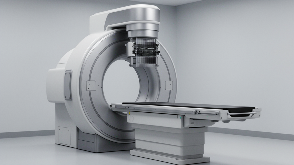
Researchers have demonstrated that the SIMIND Monte Carlo simulation code accurately reproduces the fundamental photon interaction characteristics of clinical single photon emission computed tomography (SPECT) gamma cameras. Published in EJNMMI Physics, the study provides a comprehensive validation of the simulation tool against experimental measurements from two distinct clinical systems: the Siemens E.Cam and the GE Infinia. The primary objective was to assess the reliability of SIMIND in modeling energy spectra, energy resolution, sensitivity, and image profiles, thereby establishing a robust computational framework for optimizing quantitative SPECT protocols and guiding future system design improvements.
The study employed a dual-system evaluation approach, utilizing standardized phantoms and radioactive sources to compare simulated and experimental performance metrics. For energy resolution and sensitivity assessments, an isotropic Technetium-99m (99mTc) point source emitting 140 keV photons was used. Spatial resolution was evaluated using a Cobalt-57 (57Co) flood source placed on a bar phantom for experimental measurements, while a QC Pie Phantom was utilized in the simulations to qualitatively assess resolution limits. The SIMIND code, version 6.1.2, was configured with manufacturer-specified detector geometries and material properties to ensure physical accuracy in modeling radiation detection from photon interaction to signal generation.
A key methodological contribution of this work is the development of a custom algorithm for automatic energy spectrum decomposition. This algorithm was designed to identify and characterize key spectral components, including the photopeak, Compton edge, iodine escape peak, and lead X-ray peaks. By implementing this peak identification algorithm in C++, the researchers ensured uniformity in performance validation across both experimental and simulated data. The algorithm applies smoothing filters to reduce statistical fluctuations before identifying local maxima, allowing for precise calculation of the full width at half maximum (FWHM) and energy resolution. This approach addresses a gap in previous studies that often lacked comprehensive quantitative characterization of secondary spectral peaks critical for spectral fidelity.
The results for the Siemens E.Cam system showed excellent agreement between simulated and experimental measurements. Energy resolution differences were observed to be less than 4%, with specific discrepancies of 0.4% or less between simulated and experimental FWHM values for the 140 keV photopeak. The experimental FWHM was 14.58 keV, corresponding to a relative energy resolution of 10.42%, while Monte Carlo simulations yielded FWHM values of 14.05 keV and 14.11 keV for collimated and uncollimated configurations, respectively. These values align closely with the manufacturer’s specified resolution of 9.9% at 140 keV, confirming proper system calibration and simulation parameterization.
Sensitivity measurements also demonstrated close agreement for the Siemens E.Cam, with an experimental sensitivity of 136.83 counts per second per megabecquerel (cps/MBq) compared to a simulated value of 138.35 cps/MBq. This represents a difference of only 1.1%, which falls within the bounds of acceptable experimental error. The close correspondence in sensitivity validates the accuracy of the detector modeling, particularly regarding the precise incorporation of collimator geometry and detector material properties into the simulation. The study notes that sensitivity is significantly affected by these parameters, and the minimal variation highlights the robustness of the SIMIND model for this specific camera system.
For the GE Infinia system, the study similarly observed strong agreement in energy resolution and sensitivity metrics. The experimental FWHM for the 140 keV photopeak was 13.44 keV, corresponding to an energy resolution of 9.80%, while simulations predicted a slightly broader resolution of 13.84 keV (9.60%). The difference between simulation and experiment was approximately 2.0%. Sensitivity values for the GE Infinia showed an experimental result of 107.67 cps/MBq versus a simulated 108.91 cps/MBq, a difference of 1.15%. These results confirm that the SIMIND code can accurately replicate the photon detection characteristics of the Low Energy General Purpose (LEGP) collimator used in the GE Infinia system.
Image profile analysis revealed distinct differences between the two systems. For the Siemens E.Cam, 2D and 3D image profiles showed strong agreement between simulation and experiment, with FWHM values ranging from 4 to 4.5 pixels. This close correspondence indicates that the simulation accurately represents spatial response attributes, including intrinsic resolution and collimator blur. In contrast, the GE Infinia results revealed a system-specific discrepancy. Experimental data exhibited sub-optimal uniformity and linearity, resulting in broadened point-source profiles without a distinct sharp peak. The simulated profiles, while aligning with theoretical expectations, replicated the observed “star effect” artifact due to the inherent limitations of the clinical GE Infinia system installed at the facility.
The discrepancy in the GE Infinia image profiles is attributed to the actual clinical condition of the installed camera, including compromised detector uniformity and electronic noise, rather than a failure of the SIMIND model. SIMIND models an idealized, properly calibrated SPECT system based on defined physical and geometrical parameters. Therefore, local hardware issues, photomultiplier tube gain variations, or imperfect calibration in the clinical detector may not be fully reproduced unless specifically characterized and introduced into the simulation. Despite these spatial differences, the energy spectrum characteristics and sensitivity measurements for the GE Infinia remained in close agreement with the Monte Carlo simulations, confirming the accuracy of the fundamental photon interaction modeling.
Spatial resolution assessments further highlighted system-specific capabilities. The Siemens E.Cam demonstrated a spatial resolution ranging between 3.5 mm and 4.8 mm, based on bar and pie phantom results. The pie phantom showed all rods distinctly visible, with the smallest rod diameter being 4.8 mm, while the bar phantom indicated a resolution of approximately 3.5 mm. In contrast, the GE Infinia system showed a markedly worse experimental resolution of approximately 1 cm, corresponding to the separation between the four quadrants of the bar phantom. This difference underscores the impact of detector age and calibration status on spatial performance, with the Siemens E.Cam aligning with the anticipated performance of an adequately operating Low Energy All Purpose (LEAP) collimated system.
The custom peak detection algorithm successfully identified all principal spectral features produced by 99mTc photon interactions in the NaI(Tl) crystals. These features included the photopeak at 140 keV, the Compton edge at approximately 47 keV, the lead characteristic X-ray peak at 72–75 keV, the backscatter peak at 90 keV, and the iodine escape peak at 112 keV. The differences between simulated and measured peak positions were below 3% for both systems, demonstrating good spectral fidelity. This automated identification of secondary peaks, such as the backscatter and iodine escape peaks, which were not clearly separated in previous SIMIND-based analyses, represents a significant advancement in the quantitative characterization of detector performance.
Comparison with previously published Monte Carlo validation studies further supports the reliability of the proposed framework. The intrinsic energy resolution values obtained in this study are consistent with reported values for clinical NaI(Tl)-based SPECT systems, falling within the expected 9.5–10.5% range. The study notes that slight differences between studies may arise from variations in detector configuration, source size, and specific energy-resolution modeling approaches. The proximity of data points to the reference line in comparisons across different SPECT systems and collimator configurations indicates good agreement between simulated and experimentally measured detector energy resolutions.
The findings confirm that SIMIND provides a robust framework for optimizing quantitative SPECT protocols, particularly for well-calibrated systems. The integration of the peak detection algorithm with sensitivity, spatial resolution, and image-profile evaluations offers a useful methodology for SPECT performance characterization. This framework not only validates the agreement between simulation and measurement for well-modeled detector responses but also helps identify deviations between ideal Monte Carlo predictions and the actual performance of clinical gamma camera systems. The validation represents an essential step before the simulation can be used in more complicated studies of optimizing performance characteristics and enhancing image quality in nuclear medicine diagnostics.
Subject of Research: Validation of SIMIND Monte Carlo simulation code for SPECT gamma camera performance evaluation
Article Title: Monte Carlo simulation and experimental validation of SPECT systems via energy spectrum decomposition and image profile analysis
Article References: Peng, Z., Wasaye, M. A., Siddique, I., Wali, F., & Saeed, M. (2026). Monte Carlo simulation and experimental validation of SPECT systems via energy spectrum decomposition and image profile analysis. EJNMMI Physics, 13(1), Article 90. https://doi.org/10.1186/s40658-026-00929-6
Image Credits: AI Generated
DOI: 10.1186/s40658-026-00929-6
Keywords: SPECT, Monte Carlo simulation, SIMIND, gamma camera, energy resolution, nuclear medicine, Siemens E.Cam, GE Infinia, image profile analysis, detector validation, Monte, Carlo
News Source: Denise Maddox. (November 8, 2026). SIMIND Simulation Validates SPECT Camera Performance Metrics. Scienmag.