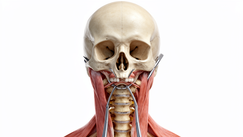
Surgical navigation around the anterior craniovertebral junction and foramen magnum presents significant technical challenges due to the dense surrounding neurovascular anatomy and constricted anatomical corridors. A laboratory investigation published in Acta Neurochirurgica provides quantitative morphometric data evaluating surgical freedom, operational angles, and volumetric exposure across standard access routes. Researchers dissected five latex-injected human cadaveric specimens to rigorously compare the transcranial far-lateral transcondylar approach, the endoscopic endonasal far-medial approach, and a combined staging of both corridors. The objective was to characterize the spatial exposure boundaries and maneuverability profiles that define these surgical interventions within a controlled laboratory setting.
The anatomical territory of the lower third of the clivus extends inferiorly to the foramen magnum, bounded laterally by the occipital condyles and the primary cervical vertebral articulation. Within this compact corridor reside vital neurovascular structures, including the vertebral arteries, anterior and posterior spinal arteries, the spinal root of the accessory nerve, and lower cranial nerves spanning nine through twelve. The hypoglossal nerve traverses anterolaterally through the subarachnoid space toward the hypoglossal canal between the occipital condyle and the jugular foramen. Additionally, the vertebral artery undergoes a transition from its third segment to its intradural fourth segment near the foramen magnum, ascending to establish the basilar trunk while typically providing the posterior inferior cerebellar artery.
To directly assess instrument handling within these corridors, the investigators quantified surgical freedom and specific angular trajectories using image-guided neuronavigation systems and three-dimensional computed tomography segmentation. Across the five dissected heads, dissections were completed unilaterally via the endoscopic far-medial approach and the contralateral far-lateral approach, followed by combined analysis. The far-lateral approach utilized microsurgical instrumentation under an operative microscope, while the endoscopic far-medial corridor employed combinations of zero-degree, thirty-degree, and forty-five-degree rigid endoscopes. Institutional cadaveric protocols governed the anatomical procedures, which incorporated pre-dissection thin-slice computed tomography scans to support registration and coordinate measurement.
Surgical freedom was experimentally defined as the allowable operating area at the proximal end of a nineteen-millimeter probe while its distal tip remained anchored at specific anatomical targets. Horizontal and vertical angles of attack were derived from the boundaries of lateral, medial, rostral, and caudal instrument travel. When measuring overall maneuverability, the far-lateral transcondylar approach provided a total surgical freedom of 363 square millimeters, with an observed range of 361 to 368 square millimeters. In contrast, the endoscopic endonasal far-medial approach yielded a total surgical freedom of 6.8 square millimeters, ranging from 6.7 to 7.6 square millimeters, a difference that reached statistical significance with a p-value of 0.01 under Wilcoxon rank-sum testing.
Measurements of instrument trajectory demonstrated a corresponding pattern across the investigated corridors. The overall horizontal angle of attack achieved through the far-lateral approach averaged 99 degrees, with a range between 98 and 101 degrees, compared to 8.1 degrees, ranging from 6.9 to 11.2 degrees, for the endoscopic far-medial approach. Similarly, the vertical angle of attack measured 78 degrees, spanning 78 to 89 degrees, in the far-lateral dissections, whereas the endoscopic far-medial corridor afforded 15.4 degrees, spanning 13.9 to 17.9 degrees. These differences across primary angular dimensions were statistically significant at a p-value of 0.01, highlighting broader instrumental reach through the posterolateral pathway.
Targeted assessments at defined anatomical structures reflected these global maneuverability differences. At the hypoglossal canal, the far-lateral approach yielded a surgical freedom of 138 square millimeters compared to 1.2 square millimeters for the endoscopic approach. The horizontal angle of attack at this landmark was 41 degrees for the far-lateral versus 4.5 degrees for the endoscopic approach, and the vertical angle of attack was 46 degrees versus 4.2 degrees. At the jugular foramen, the far-lateral route achieved 101 square millimeters of surgical freedom, a horizontal angle of 52 degrees, and a vertical angle of 41 degrees, whereas the endoscopic approach produced 1.5 square millimeters of freedom, a 4.8-degree horizontal angle, and a 4.5-degree vertical angle. At the vertebrobasilar junction, surgical freedom reached 46 square millimeters in the far-lateral corridor compared to 1.3 square millimeters endoscopically.
In contrast to the divergent maneuverability values, linear cranial nerve exposure along specific segments was comparable between the two standalone techniques. Intradural exposure of the hypoglossal nerve measured an average of 1.0 centimeter in the far-lateral approach and 1.1 centimeters in the endoscopic far-medial approach, showing no statistically significant variation with a p-value of 0.83. Glossopharyngeal nerve exposure likewise averaged 1.3 centimeters in both approaches, yielding a p-value of 0.58. However, bony resection around the perimeter differed, as the degree of foramen magnum margin removal measured 129 degrees in the far-lateral dissections compared to 45 degrees in the endoscopic dissections, representing a statistically significant difference with a p-value of 0.01.
Volumetric analysis based on three-dimensional manual segmentation revealed distinct spatial distributions between corridors. Segmented corridors were subdivided relative to the cisternal segments of the lower cranial and hypoglossal nerves into anteromedial and posterolateral compartments. The endoscopic endonasal far-medial approach yielded the lowest mean total volume at 5.53 cubic centimeters, which was predominantly situated in the anteromedial compartment at 4.70 cubic centimeters, with minimal posterolateral volume measuring 0.83 cubic centimeters. Conversely, the far-lateral approach generated a significantly larger mean total exposure of 15.12 cubic centimeters, dominated by 12.60 cubic centimeters of posterolateral space and 2.52 cubic centimeters of anteromedial access.
Integrating both trajectories into a combined approach established the largest overall surgical volume, measuring an average of 18.76 cubic centimeters. This staged model encompassed 12.90 cubic centimeters of posterolateral volume alongside 5.86 cubic centimeters of anteromedial volume. While combining approaches significantly increased total and posterolateral volume when compared to the endoscopic approach alone, the comparison against the isolated far-lateral approach showed no significant difference in posterolateral exposure. The combined procedure achieved an increase in anteromedial volume relative to the standalone far-lateral approach, recording a p-value of 0.013, although the practical implementation of combined corridors requires separate patient repositioning, extended anesthesia, and staged operative intervals.
The study authors noted inherent methodological constraints, emphasizing that the measurements reflect anatomically intact, non-pathological cadaveric tissues. Clinical lesions, such as meningiomas or chordomas, often distort regional landmarks, erode bone, or displace neurovascular pathways, which can alter operative corridors in vivo. Despite these limitations, the quantitative findings establish baseline parameters indicating that the far-lateral corridor provides superior surgical freedom and posterolateral exposure, whereas the endoscopic far-medial route provides a focused, ventral corridor primarily restricted to the anteromedial compartment.
The surgical procedures followed precise multi-stage protocols to establish access. In the far-lateral approach, dissection progressed through three distinct phases: muscular dissection utilizing an interfascial plane to safely expose the vertebral artery within the suboccipital triangle, extradural dissection encompassing a lateral suboccipital craniotomy, a first cervical hemilaminectomy, and removal of the lateral foramen magnum margin, followed by drilling the posteromedial occipital condyle using a three-millimeter diamond burr to reach the hypoglossal canal, jugular tubercle, and premedullary region. Conversely, the endoscopic far-medial approach operated as a transcondylar and transjugular tubercle extension of a transclival trajectory. This corridor commenced with a posterior septectomy and inferior clivectomy, extending laterally toward the occipital condyle before resecting the atlanto-occipital joint capsule to reveal the supracondylar groove. Subsequent bone removal along the inferolateral clivus exposed the anterior cortical boundary of the hypoglossal canal, effectively partitioning the lateral clival corridor into a superior tubercular compartment and an inferior condylar compartment to permit intradural visualization of the ninth through twelfth cranial nerves.
These quantitative anatomical distinctions correspond directly to technical demands and risks documented across clinical literature. Transcranial far-lateral techniques have traditionally served as workhorse corridors for posterolateral craniovertebral lesions, including lower cranial nerve schwannomas and lateral foramen magnum meningiomas. Previous comparative cadaveric studies noted that while the far-lateral route commands seventy-four percent of dorsolateral brainstem exposure and permits instrument passage across lower cranial nerves, the endoscopic far-medial corridor provides superior exposure of the ventromedial brainstem. Clinically, endonasal routes have been applied to anterior pathologies such as clival chordomas, chondrosarcomas, and extracranial hypoglossal schwannomas without requiring external incisions or suboccipital muscle reflection. However, the endoscopic approach involves a steep learning curve and carries well-recognized clinical hazards, most notably higher rates of postoperative cerebrospinal fluid leakage and an elevated potential for cranial nerve injury when attempting lateral resection near the occipital condyle.
To synthesize these findings into operative decision-making, the authors outlined an anatomy-driven selection framework based on lesion localization and neurovascular displacement. Pathologies located strictly anterior to the brainstem frequently displace neural and vascular structures posteriorly, restricting the lateral-to-medial working corridor of the far-lateral approach and making the ventral endoscopic trajectory preferable. For instance, small tumors situated anterior to neurovascular bundles, classified as Type 3 lesions, align well with direct endoscopic resection. In contrast, lesions centered laterally or posterolaterally, or those extending beyond the medial jugular tubercle, remain better managed through far-lateral exposures, where condylar drilling can broaden the operative field. For extensive Type 4 tumors that encase vital vascular trunks or span multiple compartments across the midline, the quantitative data justify considering a staged combined approach despite the procedural demands of dual positioning.
Subject of Research: Quantitative comparison of skull base surgical corridors to the foramen magnum
Article Title: Comparative analysis of endoscopic endonasal far-medial, far-lateral transcondylar and combined approaches to the foramen magnum region: volumetric analysis and quantitative insights on surgical exposure and maneuverability
Article References: Blue, R., Zhou, Y., Bond, K., Ajmera, S., Alvarez, R., Moseley, R., Hosokawa, P., & Youssef, A. S. (2026). Comparative analysis of endoscopic endonasal far-medial, far-lateral transcondylar and combined approaches to the foramen magnum region: volumetric analysis and quantitative insights on surgical exposure and maneuverability. Acta Neurochirurgica, 168(1), Article 227. https://doi.org/10.1007/s00701-026-07039-3
Image Credits: AI Generated
DOI: 10.1007/s00701-026-07039-3
Keywords: Neurosurgery, Skull base surgery, Far-lateral approach, Endoscopic endonasal approach, Foramen magnum, Craniovertebral junction, Surgical maneuverability, Cadaver study, Comparative, analysis, endoscopic, endonasal
News Source: Ophelia Keating. (November 8, 2026). Cadaver Study Compares Surgical Approaches to Craniovertebral Junction Corridors. Scienmag.