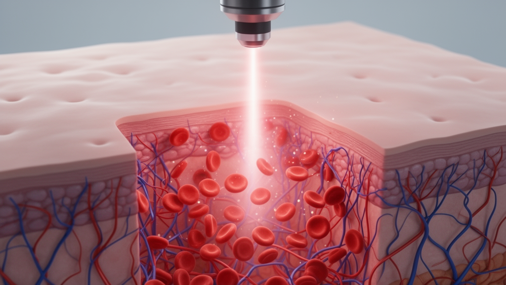
Researchers have developed a novel computational approach that enhances the ability of laser speckle contrast imaging to measure blood flow velocity independently of vessel depth. Published in the journal Frontiers of Optoelectronics, the study presents a method that combines laser speckle contrast imaging with principal component analysis and entropy analysis to separate static and dynamic components of the speckle signal. This technique aims to overcome a significant limitation of conventional laser speckle imaging, where the presence of static scattering layers above or below blood vessels reduces image contrast and resolution, thereby complicating accurate blood flow assessment.
The study, led by Yu. Surkov and colleagues, utilized optical phantoms and in vivo experiments on a laboratory mouse ear to validate the proposed method. The optical phantoms were constructed using epoxy resin containing titanium dioxide microparticles as scatterers, with a glass capillary embedded at an angle to simulate vessels at varying depths ranging from 0.6 to 2 mm. A 3% aqueous solution of intralipid was used to model the scattering properties of blood, with flow rates adjusted using a syringe pump to simulate different linear velocities. The experimental setup included a single-mode He–Ne laser with a wavelength of 632.8 nm and a monochrome CMOS camera equipped with a micro-objective to record raw speckle images.
Principal component analysis was applied to filter the speckle signal, separating it into static and dynamic components. The static component, representing signals from stationary tissue structures, was identified using the Guttman-Kaiser criterion, while the dynamic component, reflecting motion from moving scatterers such as blood cells, was derived by subtracting the static component from the original signal. This separation allows for a more accurate assessment of blood flow dynamics by minimizing the influence of static scattering layers that can otherwise distort the speckle pattern and lead to incorrect interpretations of blood flow changes.
The results demonstrated that the proposed method significantly improves the visualization of subsurface blood vessels by enhancing image contrast and resolution. In the optical phantom experiments, the method showed a substantial increase in the sensitivity to blood flow velocity within the physiologic range of 0.98 to 19.66 mm/s, while minimizing sensitivity to vessel depth. For laser speckle contrast imaging methods combined with principal component analysis, the Pearson correlation coefficient between the speckle flow index and linear fluid velocity increased across the entire range of vessel depths, from 0.63 to 1.95 mm.
Specifically, for transmissive-detected laser speckle contrast imaging with principal component analysis, the average correlation coefficient increased from 0.80 to 0.95, representing an 18.7% improvement. For reflective-detected laser speckle contrast imaging, the correlation coefficient increased from 0.78 to 0.92, a 17.9% increase. The highest correlation coefficient was observed for spatiotemporal laser speckle contrast imaging combined with principal component analysis, reaching 0.99 at a depth of 0.63 mm and maintaining a value of 0.96 at a depth of 1.95 mm. These findings indicate that the method provides a more reliable and consistent measurement of blood flow velocity regardless of the depth of the vessel being imaged.
In contrast, laser speckle entropy imaging combined with principal component analysis did not show significant improvements in correlation with linear velocity. However, laser speckle entropy imaging without principal component analysis demonstrated a higher correlation with linear velocity compared to laser speckle contrast imaging methods without the filtering technique.
The in vivo experiments on a laboratory mouse ear further confirmed the effectiveness of the proposed approach. Images obtained using the method with principal component analysis showed significantly more detailed and contrasted vascular networks compared to traditional laser speckle imaging. The dynamic component images revealed clearer vessel structures with reduced background noise and artifacts, allowing for better visualization of blood flow in the microvasculature. The study noted that the transmissive detection mode produced sharper and more contrasted images than the reflective detection mode, although both benefited from the application of principal component analysis.
The ability to assess blood flow velocity independently of vessel depth is a critical advancement for non-invasive biomedical imaging. Conventional laser speckle imaging techniques often struggle to provide accurate measurements for deeper vessels due to the attenuation and distortion of the speckle signal by overlying tissue. By effectively separating the static and dynamic components of the speckle signal, the new method mitigates these issues, offering a more robust tool for monitoring blood flow in various biological tissues. This capability is particularly valuable in clinical diagnostics and biomedical research, where high precision in blood flow monitoring is essential for assessing tissue viability and detecting perfusion issues.
The study also highlighted the importance of the detection mode in laser speckle imaging. Transmissive-detected laser speckle contrast imaging, which relies on forward-scattered light, was found to be more effective for deep visualization compared to reflective-detected methods, which rely on backscattered light. This difference is attributed to the higher intensity of scattered radiation associated with the useful signal in the transmissive mode when the vessel depth exceeds 200 micrometers. The findings suggest that the choice of detection mode can significantly impact the quality of the imaging and the accuracy of the blood flow measurements, especially in the presence of thick scattering layers.
Limitations of the study include the use of a single mouse ear for the in vivo demonstration, which was chosen to focus on the reproducibility and reliability of the method rather than biological variability. The authors noted that future studies should expand the sample size to investigate biological variability and further validate the method in more complex physiological environments. Additionally, the method’s performance in different tissue types and under various pathological conditions remains to be fully explored. Despite these limitations, the study provides a strong foundation for the development of advanced laser speckle imaging techniques that can enhance the accuracy and reliability of blood flow assessment in biomedical applications.
The proposed approach holds great potential for advancing the use of laser speckle imaging in clinical settings, where non-invasive and real-time monitoring of blood flow is crucial. By overcoming the depth-dependence issue, the method could enable more precise diagnostics and treatment planning for conditions affecting microcirculation, such as stroke, tissue necrosis, and surgical flap viability. The integration of principal component analysis and entropy analysis into laser speckle imaging represents a significant step forward in the field of optical biomedical imaging, offering new possibilities for the visualization and quantification of blood flow in scattering tissues.
As the field of laser speckle imaging continues to evolve, the development of such computational methods will be essential for improving the clinical utility of these techniques. The ability to accurately measure blood flow velocity independent of vessel depth opens up new avenues for research and clinical application, potentially leading to better patient outcomes and a deeper understanding of microcirculatory dynamics. The study underscores the importance of innovative signal processing techniques in enhancing the performance of existing imaging modalities, paving the way for more advanced and reliable tools in biomedical imaging.
Subject of Research: Laser speckle contrast imaging with principal component and entropy analysis: a novel approach for depth-independent blood flow assessment
Article Title: Laser speckle contrast imaging with principal component and entropy analysis: a novel approach for depth-independent blood flow assessment
Article References: Surkov, Y., Timoshina, P., Serebryakova, I., Stavtcev, D., Kozlov, I., Piavchenko, G., Meglinski, I., Konovalov, A., Telyshev, D., Kuznetcov, S., Genina, E., & Tuchin, V. (2025). Laser speckle contrast imaging with principal component and entropy analysis: a novel approach for depth-independent blood flow assessment. Frontiers of Optoelectronics, 18(1), Article 1. https://doi.org/10.1007/s12200-024-00143-1
Image Credits: AI Generated
DOI: 10.1007/s12200-024-00143-1
Keywords: Laser, speckle, contrast, imaging, principal, component, entropy, analysis, novel, approach, depth-independent, blood
News Source: Denise Maddox. (November 8, 2026). New Laser Imaging Method Measures Blood Flow Depth Independently. Scienmag.