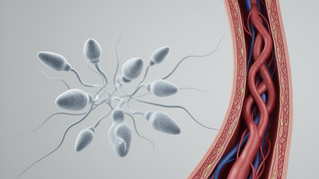
In a cross-sectional investigation examining semen samples from 468 men undergoing evaluations at an infertility clinic, researchers found that specific sperm structural defects and bilateral varicoceles were significantly correlated with elevated sperm DNA fragmentation. Elevated DNA fragmentation index values, which reflect impaired genomic integrity in spermatozoa, showed notable associations with micro- and partial-head sperm abnormalities as well as marked reductions in conventional semen parameters including sperm concentration, progressive motility, and overall normal morphology.
Empirical evidence indicates that high levels of sperm DNA fragmentation negatively influence reproductive outcomes across both natural conception and assisted reproductive technologies, including in vitro fertilization and intracytoplasmic sperm injection. Infertility affects approximately 17 percent of individuals during their reproductive lifespan and contributes to nearly half of all couple infertility cases, either as the sole factor or combined with female factors. Although standard semen analyses evaluating sperm count, motility, and morphology provide baseline diagnostic information, many men with seemingly normal results still experience infertility, highlighting the clinical necessity of assessing sperm DNA integrity.
Despite the recognized role of sperm DNA integrity in embryonic development and blastocyst quality, routine testing often presents an additional financial cost to patients, and detailed clinical guidelines indicating which patient groups should undergo targeted testing based on initial semen profiles have remained limited. To address this diagnostic gap, the study aimed to evaluate how standard semen classifications and detailed sperm morphology subcategories correspond to elevated fragmentation levels, establishing practical criteria to guide selective testing in male infertility evaluations.
The study protocol evaluated 468 men aged 20 to 55 years who were referred to the infertility center of Taleghani Hospital in Tehran, Iran. Semen specimens were collected after three to five days of sexual abstinence and allowed to liquefy before undergoing manual evaluation in accordance with World Health Organization guidelines. All 468 collected samples fulfilled the inclusion criteria and were stratified into three distinct categories based on their measured DNA fragmentation index: a low fragmentation cohort with index values under 20 percent comprising 150 men, an intermediate cohort with values between 20 and 25 percent comprising 150 men, and a high fragmentation cohort exceeding 25 percent comprising 168 men.
Morphological assessments were conducted using bright-field microscopy under oil immersion at 1000-times magnification following Diff-Quik staining, scoring 200 spermatozoa per specimen. To categorize structural anomalies, researchers applied a strict hierarchical registration system that registered only one defect per cell, prioritizing head defects over neck defects, which were prioritized over tail defects. Morphological anomalies recorded included large, pyriform, tapered, pin, micro- or partial-head, globozoospermic, amorphous, and vacuolated structures, alongside asymmetrical insertions, residual cytoplasmic droplets, and coiled or stump tails.
Sperm DNA integrity was assessed using the sperm chromatin dispersion assay, which quantifies the presence of nuclear chromatin dispersion halos following nuclear protein extraction and DNA denaturation, with absent halos denoting fragmented DNA across 300 examined cells per sample. Concurrently, sperm chromatin maturation was determined through aniline blue staining via the sperm chromatin maturation assay, scoring 200 spermatozoa per smear to calculate the proportion of cells with immature chromatin indicated by blue-stained nuclei. The ethical committee of the Faculty of Medicine at Shahed University approved the study protocol, and all enrolled participants provided written informed consent.
Methodological reproducibility was validated on a randomly selected subset of 29 slides evaluated blindly by an experienced embryologist and re-scored after an interval of at least two weeks, alongside independent scoring by a second trained evaluator. Continuous agreement for DNA fragmentation measurements demonstrated an intraclass correlation coefficient of 0.968 for intra-observer testing and 0.994 for inter-observer comparison. Categorical agreement using the 25 percent fragmentation cut-off yielded Cohen’s kappa coefficients ranging from 0.84 to 1.00, demonstrating substantial to almost-perfect analytical consistency.
Analysis of participant baseline characteristics revealed that mean age and duration of infertility did not differ significantly across the low, intermediate, and high fragmentation cohorts, nor did lifestyle factors such as smoking history. However, clinical presentation of bilateral varicocele was significantly more prevalent among men categorized in the high fragmentation group compared to those with lower fragmentation values. Semen volume remained statistically comparable across all three groups.
Conventional semen metrics exhibited pronounced differences across the fragmentation categories, with the high fragmentation group displaying significantly lower sperm concentrations, reduced total motility, and diminished progressive motility compared to samples in lower index brackets. Normal morphological forms were similarly reduced at higher fragmentation levels, while the proportion of immature spermatozoa demonstrated a direct association with escalating fragmentation indices. Semen categorization revealed a statistically significant low-power correlation between DNA fragmentation and asthenozoospermia combined with oligozoospermia or teratozoospermia.
Detailed subcategorization of structural defects demonstrated that micro- and partial-head abnormalities exhibited the strongest positive correlation with elevated fragmentation, presenting an intermediate correlation coefficient of 0.40. Other head variations, such as tapered and amorphous configurations, displayed elevated frequencies in samples with high fragmentation, but did not reach statistical significance across all comparisons. In specimens evaluated against a 24 percent chromatin immaturity threshold, tapered head morphology was more frequently observed in samples with higher fragmentation indices compared to more mature profiles.
The investigators outlined several biological mechanisms that likely account for the observed link between sperm head dysmorphology and impaired genomic integrity during spermatogenesis. Failure of morphogenesis during the early spermatid phase can impede the repair of physiological DNA strand breaks, while defective spermiogenesis disrupts nuclear reshaping and chromatin condensation, wherein abnormal protamine-DNA complex assembly compromises structural stability. Furthermore, spermatozoa with incomplete maturation produce excessive levels of reactive oxygen species, generating oxidative stress that induces both structural head abnormalities and extensive oxidative DNA strand fragmentation.
The study authors highlighted several methodological limitations, noting that the cross-sectional design and single-center sample size of 468 men require validation in larger prospective cohorts before establishing definitive diagnostic thresholds. Additionally, while the sperm chromatin dispersion test provides consistent analytical metrics, it may not represent the sole clinical gold standard across all laboratory environments. The researchers concluded that targeted assessment of sperm DNA integrity is particularly justified in patients exhibiting bilateral varicoceles, combined oligo-astheno-teratozoospermia, or elevated frequencies of micro- and partial-head sperm defects, offering an evidence-based approach to refining male infertility diagnostics.
Beyond overall correlation coefficients, the analysis of analytical variability provided deeper insight into the precision of the scoring procedures. In the random subset of 29 slides re-evaluated to assess observer consistency, the mean absolute percent difference between paired scores was 11.08 percent between the baseline score and the first re-score, 4.52 percent between baseline and the second observer, and 11.35 percent between the two independent evaluators. These reproducibility metrics reinforced the reliability of the bright-field microscopic scoring of chromatin dispersion halos, wherein intact spermatozoa display distinct outer halos of dispersed DNA loops while cells with fragmented DNA completely lack evident halos across the 300 examined cells.
Interestingly, when examining samples categorized as normozoospermic, the investigators found no significant differences across the low, intermediate, and high fragmentation cohorts. This finding underscores that while standard semen profiles may occasionally mask underlying genomic damage, substantial elevations in fragmentation indices cluster primarily among specimens displaying compromised motility, count, or morphology. The authors noted that prior research links abnormal sperm metrics to reduced antioxidant concentrations and elevated oxidative stress markers in seminal plasma. Such oxidative imbalances not only induce DNA strand breaks but also correlate with chromatin decondensation and higher rates of immature sperm displaying protamine deficiency.
The study also situated the observed link between bilateral varicoceles and elevated fragmentation within broader clinical evidence. Men presenting with varicoceles consistently exhibit higher rates of immature sperm and chromatin abnormalities compared to fertile controls, a phenomenon observed across both normozoospermic and oligoasthenoteratozoospermic cohorts. The underlying pathogenesis centers on an imbalance between excessive reactive oxygen species generation and insufficient antioxidant protection. Notably, prior studies cited by the researchers have demonstrated that surgical intervention via varicocelectomy can improve sperm DNA integrity, leading to significant decreases in fragmentation indices and improvements in chromatin compaction post-surgery. Oxidative stress further correlates positively with chromatin decondensation in these patients.
The morphological examination also highlighted the diagnostic relevance of specific head defects beyond simple size variations. Structural abnormalities detailed in the methodology, including amorphous forms, post-acrosomal vacuoles, and disproportionate acrosomal areas covering less than 40 percent or more than 70 percent of the head area, reflect disrupted nuclear development. During spermiogenesis, significant nuclear rearrangements, chromatin condensation, and nuclear reshaping take place within the sperm nucleus. The shaping of sperm heads depends on specific patterns of DNA-protein complex assembly established during condensation, where protamine deficiency contributes directly to DNA instability and diminished quality. When this developmental process is impaired, immature spermatozoa generate higher levels of reactive oxygen species, compounding structural defects with oxidative DNA damage.
To evaluate data distributions across all parameters, the researchers tested for normality using the Shapiro-Wilk test, directing the subsequent application of analysis of variance and Kruskal-Wallis tests, followed by Tukey post-hoc analyses and Pearson correlations evaluated at a significance threshold of p less than 0.05. Crucially, the authors emphasized that because the observed correlations between fragmentation and clinical features, including bilateral varicoceles, combined asthenozoospermia, and micro- or partial-head defects, were of low-to-moderate strength, they should be interpreted strictly as biological associations rather than definitive predictive thresholds. Consequently, the investigators concluded that larger prospective studies remain essential to determine whether specific fragmentation cut-offs can be validated for clinical decision-making.
Subject of Research: Predictors of elevated sperm DNA fragmentation identified through semen and morphology analysis
Article Title: Predictors of elevated sperm DNA fragmentation: a morphology-based approach to semen analysis
Article References: Kazemi, M., Moradi, A., Bayat, F., Salehpour, S., Niakan, S., & Nazarian, H. (2025). Predictors of elevated sperm DNA fragmentation: a morphology-based approach to semen analysis. Basic and Clinical Andrology, 35(1), Article 48. https://doi.org/10.1186/s12610-025-00297-8
Image Credits: AI Generated
DOI: 10.1186/s12610-025-00297-8
Keywords: sperm DNA fragmentation, semen analysis, male infertility, sperm morphology, varicocele, assisted reproductive technology, spermiogenesis, andrology, Predictors, elevated, sperm, fragmentation
News Source: Ophelia Keating. (November 8, 2026). Sperm Head Defects and Bilateral Varicocele Predict Elevated Semen DNA Fragmentation. Scienmag.