A European research collaboration led by scientists from Heinrich Heine University Düsseldorf, the ELI Beamlines research facility, and the University of Strathclyde has demonstrated through computer model simulations that compact laser-plasma particle accelerators can provide practical access to soft X-ray radiation in the specialized “water window.” Reporting their findings in the peer-reviewed scientific journal Nature Communications, the team showed that laser-driven accelerator technology can produce the high-brightness radiation historically restricted to massive research installations. Soft X-rays occupying this spectrum have remained technically difficult to generate at sufficient intensity and beam quality without relying on vast accelerator infrastructure. The newly modeled approach demonstrates that laser-plasma systems can generate the necessary electron beams to drive free-electron laser emissions within a compact physical footprint, opening new avenues for structural analysis in the life sciences.
The water window designates electromagnetic radiation with wavelengths spanning approximately 2.3 to 4.4 nanometres, an energy band that offers exceptional advantages for inspecting biological specimens. Because biological systems consist predominantly of water while biological molecules are largely composed of elements such as carbon and nitrogen, this specific radiation regime permits high-contrast imaging of organic structures. Water remains transparent to soft X-rays within this wavelength interval, whereas carbon and nitrogen atoms absorb the radiation strongly, enabling scientists to examine biological samples directly within their natural, aqueous environments at high resolution. Professor Dr Bernhard Hidding, leader of the strong-field laser plasma physics group at Heinrich Heine University Düsseldorf and corresponding author of the study, noted that the scientific community has historically depended on kilometre-long free-electron laser systems driven by conventional electron accelerators to generate sufficiently bright radiation for such biological examinations. Together with the ELI Beamlines facility in Dolní Břežany in Czechia and the University of Strathclyde in Scotland, the researchers designed an alternative pathway utilizing a compact electron accelerator based on laser-plasma technology, key aspects of which were developed in Düsseldorf.
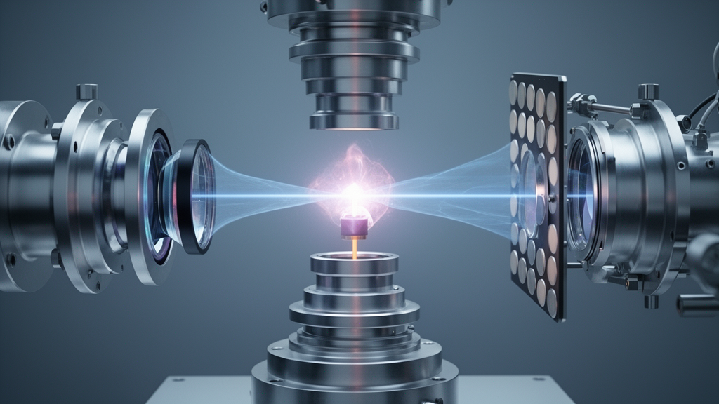
In a laser-plasma accelerator, a high-intensity laser pulse generates a plasma wave in a gas medium. The exceptionally high field intensity created within the plasma wave can accelerate electrons to nearly the speed of light across a distance of just a few centimetres. At Heinrich Heine University Düsseldorf, this acceleration concept is demonstrated by the ARCTURUS laser system, which generates intense electron beams for research and development directed toward producing X-ray flashes in the water window. To accomplish this, green laser pulses pump titanium-sapphire crystals, generating high-intensity laser pulses in the near-infrared range. In turn, these near-infrared laser pulses drive plasma waves that produce high-energy electron beams designed, in the final stage of the process, to generate intense X-ray flashes on the basis of the free-electron laser principle.
To generate the desired X-ray flashes, the high-energy electron beams are directed into magnetic undulators. An undulator is a specialized structure comprising a series of dipole magnets arranged with alternating polarity. When an electron passes through these magnetic fields, it is forced to follow a sinusoidal path. The continuous acceleration that occurs along this path causes the particles to emit synchrotron radiation, which consists of electromagnetic radiation whose wavelength can be precisely controlled via the shape and amplitude of the sinusoidal trajectory. Lily Berman, doctoral researcher in Professor Hidding’s working group at the University of Strathclyde and lead author of the study, noted that the simulations show that novel, ultra-bright electron pulses in magnetic undulators can produce X-ray flashes within just a few metres. Berman explained that this configuration converts the maximum amount of electron energy into X-ray energy, while providing a process that should be robust and flexibly adjustable.
Building on undulator technology, the system exploits the interaction between the emitted radiation and subsequent electrons traveling through the structure. The radiation that has already been emitted interacts with following electrons, bunching them into small packages. During their subsequent passage through the undulator, these bunched packages emit their synchrotron radiation in phase, meaning that all light waves oscillate at exactly the same rate. Through this avalanche mechanism, which amplifies radiation in the same manner as a conventional laser, the free-electron laser produces ultra-short, extremely bright pulses of light. These intense light pulses can be tuned across a wide range of wavelengths, providing the precise characteristics needed to probe molecular structures in biological research.
Translating these theoretical advances into practical applications depends fundamentally on the physical characteristics of the generated particle beams. Dr Fahim Habib, Principal Investigator at the ELI research facility, emphasized that whether a radiation source of this kind can function successfully as a free-electron laser depends primarily on the quality of the electron beams. The research team’s study demonstrates that a novel hybrid laser-plasma accelerator can create the necessary prerequisites for high-gain laser operation. With the simulation framework established, Dr Habib indicated that the research team will test this hybrid approach experimentally in the near future to confirm its operational viability.
The development of compact accelerator technology offers substantial implications for expanding scientific infrastructure. Professor Hidding highlighted that because such a system is very compact and can be built at a fraction of the cost of conventional particle accelerators, it could be sited directly at universities and independent research facilities, or utilized for commercial purposes. As a result, water-window X-ray imaging and spectroscopy could expand from a small number of major national laboratories into the wider research landscape. The study was conducted within the framework of a strategic partnership between Heinrich Heine University Düsseldorf and ELI Beamlines, receiving funding from the German Research Foundation, the Federal Ministry of Research, Technology and Space, and European funding programmes including the European Plasma Research Accelerator with eXcellence.
Subject of Research: Compact laser-plasma particle accelerators for water-window X-ray generation
Article Title: Compact x-ray source for research in the life sciences
Article References: Compact x-ray source for research in the life sciences. Original publication (Bibliographic details unavailable.)
Image Credits: AI Generated
DOI: Not provided
Keywords: laser-plasma accelerator, free-electron laser, water window, soft X-rays, biological imaging, Heinrich Heine University, ELI Beamlines, synchrotron radiation, Compact, x-ray, source, research
News Source: Bethany Barker. (November 8, 2026). Simulations Show Compact Laser-Plasma Source Produces Water-Window X-Rays. Scienmag.