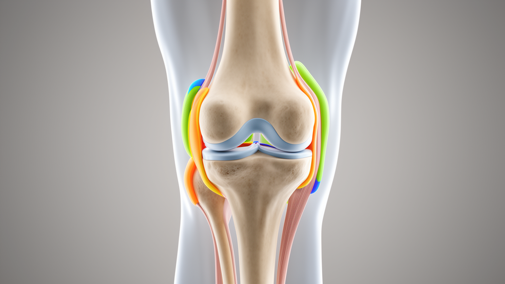
Researchers conducted a retrospective computational study to systematically compare the performance of U-Net and conditional generative adversarial networks (cGAN) for the automatic multi-tissue segmentation of knee magnetic resonance imaging (MRI). Utilizing the public SKI10 dataset, the study aimed to determine which architecture better identifies the femur, femoral cartilage, tibia, and tibial cartilage. The investigation also examined how specific loss functions, discriminator receptive fields, and normalization strategies influence model accuracy. The primary finding indicates that neither model is universally superior; instead, U-Net and cGAN exhibit complementary strengths in different aspects of segmentation quality.
The study design involved a five-class segmentation task, including the four anatomical structures and a background class. The SKI10 dataset contains 100 knee MRI volumes with manual labels. To prevent information leakage, the data were split at the volume level, with 70 volumes used for training and 30 reserved exclusively for testing. Among the training volumes, 10 were held out as a validation set for checkpoint selection. This rigorous partitioning ensured that the test set remained completely untouched during training, model selection, and hyperparameter optimization, providing an objective evaluation of model performance.
For the baseline comparison, U-Net served as the supervised model, while the cGAN framework consisted of a U-Net-style generator and a discriminator. The evaluation metrics included the Dice similarity coefficient (DSC), which measures regional overlap, and the 95th-percentile Hausdorff distance (HD95), which reflects boundary distance. The results showed that the mean DSC across the four anatomical structures was significantly higher for cGAN than for U-Net, with values of 0.8061 versus 0.8035. However, the mean HD95 was lower for cGAN than for U-Net, at 4.02 mm compared to 5.38 mm, indicating better boundary-distance control and local contour fitting for the adversarial network.
Structure-specific analysis revealed that segmentation of osseous structures was more stable than that of cartilage structures. Model differences were concentrated mainly in thin-layer tissues such as femoral and tibial cartilage. U-Net achieved a significantly higher DSC for femoral cartilage, suggesting it is better at maintaining regional overlap and cartilage continuity. In contrast, cGAN achieved significantly higher DSC for the tibia and femur, and lower HD95 across all structures. This discrepancy highlights that DSC and HD95 capture different dimensions of segmentation quality, with DSC favoring volume overlap and HD95 penalizing boundary errors.
Ablation experiments on loss functions demonstrated that L2 and Smooth L1 losses improved DSC and reduced HD95 in U-Net compared to the cross-entropy baseline. Specifically, L2 increased the mean DSC to 0.8420 and reduced HD95 to 3.89 mm. In the cGAN framework, the L1 pixel-wise loss yielded the most stable performance. These findings suggest that supervised and adversarial models respond differently to loss functions, with continuous pixel-wise constraints benefiting U-Net’s probability-map calibration, while L1 loss provides stability in the adversarial training of cGAN.
The study also investigated the effect of discriminator receptive fields on cGAN performance. Four configurations were tested: PixelGAN, PatchGAN-34, PatchGAN-70, and PatchGAN-286. PixelGAN achieved the best overall performance, with a mean DSC of 0.8276 and a mean HD95 of 3.60 mm. Larger PatchGAN receptive fields did not improve segmentation performance; in fact, PatchGAN-34 and PatchGAN-286 achieved significantly lower DSC than the baseline PatchGAN-70. This indicates that larger receptive fields do not necessarily improve the segmentation of medical label maps, possibly due to excessive morphological constraints on small target structures.
Normalization strategies were also compared, with BatchNorm and InstanceNorm evaluated in both U-Net and cGAN. BatchNorm achieved higher DSC than InstanceNorm in both models. In U-Net, BatchNorm increased mean DSC from 0.7704 to 0.8035, although InstanceNorm achieved lower HD95. In cGAN PixelGAN, BatchNorm significantly improved both DSC and HD95. These results suggest that BatchNorm is more suitable for this task, likely because it preserves within-slice intensity features that are important for distinguishing cartilage, bone, and soft tissues.
The training dynamics of the two models also differed. U-Net training was generally stable, with smooth DSC curves. cGAN training exhibited more fluctuations due to the alternating optimization between the generator and discriminator. This behavior is consistent with adversarial training characteristics and suggests that cGAN performance is sensitive to discriminator capacity and loss weighting. The study emphasizes that the cGAN advantage is associated with boundary-distance control rather than a consistent improvement in regional overlap across all tissues.
Limitations of the study include its reliance on a single predefined volume-level split of the SKI10 dataset. The results require confirmation through repeated splits, cross-validation, and external multicenter validation to assess generalizability. Additionally, the use of two-dimensional slice modeling did not fully exploit three-dimensional spatial continuity, which may affect the stable recognition of continuous thin-layer structures like cartilage. The study did not incorporate complex data augmentation or postprocessing strategies, which could further improve model stability.
In conclusion, U-Net and cGAN show complementary strengths in knee MRI multi-tissue segmentation. U-Net is more suitable for tasks emphasizing regional overlap and cartilage continuity, while cGAN, particularly in the PixelGAN configuration, is better for tasks emphasizing boundary distance and surface-shape control. These findings support task-specific selection of segmentation architectures and discriminator configurations. The study provides experimental evidence for model selection in cartilage quantification and boundary measurement, laying a foundation for future external validation and clinical quantitative applications.
The study was published in Frontiers in Molecular Biosciences, with corresponding author Minghan Li from Hubei University of Chinese Medicine and co-authors from institutions in Wuhan and Nanchang, China. The research team utilized the SKI10 dataset, which originates from the MICCAI 2010 Knee Segmentation Challenge, to ensure a standardized benchmark for evaluating segmentation algorithms. By employing a retrospective computational design, the researchers focused on isolating the specific contributions of network architecture and training parameters rather than optimizing for state-of-the-art performance on a single metric. This approach allows for a detailed dissection of how different components of deep learning models interact when processing medical imaging data, providing a controlled environment to observe the effects of adversarial training versus supervised learning in a clinical imaging context.
Preprocessing of the MRI data involved extracting two-dimensional slices along the sagittal direction from the original three-dimensional volumes. Each slice was resized to 512 by 512 pixels and standardized using z-score normalization to reduce the influence of inter-image grayscale differences. Labels were resized using nearest-neighbor interpolation to preserve categorical encoding, preventing the introduction of non-integer values that could compromise semantic segmentation accuracy. The input images were stored in float32 format, while labels were stored in uint8 format, with data organized in H5 chunks at the single-slice level to support efficient batch training on high-performance hardware. This meticulous data preparation ensures that the models are trained on consistent intensity distributions and spatial resolutions, which is critical for fair comparison between the U-Net and cGAN architectures.
The U-Net baseline model featured an encoder-decoder architecture with skip connections, containing approximately 67.0 million parameters. The discriminator was configured to receive a channel-wise concatenation of the MRI image and the segmentation map, allowing it to evaluate the structural realism and image-label consistency of the predicted outputs. This conditional discrimination mechanism enables the discriminator to learn local or global structural priors of the label map, thereby imposing morphological constraints on the generated segmentations. The use of a U-Net-style generator within the cGAN framework facilitates the simultaneous use of semantic feature extraction and spatial restoration, making it suitable for the complex anatomical structures present in knee MRI.
Training parameters were carefully standardized to ensure comparability across all experimental configurations. Both models were trained for 100 epochs using an initial learning rate of 2 times 10 to the power of negative 4. The U-Net used the AdamW optimizer with a cosine-annealing learning-rate schedule, while the cGAN used the Adam optimizer with a fixed learning rate for both the generator and discriminator. Automatic mixed precision was enabled for all models to improve computational efficiency, and no data augmentation was applied to reduce potential confounding effects on the ablation analyses. The batch sizes were set to 256 for U-Net and 128 for cGAN, with per-GPU batch sizes adjusted to fit the memory constraints of the NVIDIA A100 GPU used for training. These consistent training conditions allow the observed performance differences to be attributed to the architectural and loss-function variations rather than differences in optimization strategies.
Statistical analysis treated the MRI volume as the independent unit of observation, recognizing that slices from the same volume are not statistically independent. Paired-samples t-tests were used to compare model configurations, with the normality of paired differences examined using the Shapiro-Wilk test. The primary comparison focused on the mean DSC and HD95 across the four anatomical structures, while ablation analyses were treated as exploratory. To control the family-wise error rate, P values from the ablation analyses were corrected using the Benjamini-Hochberg false-discovery-rate procedure, and all significant comparisons remained significant after correction. Effect sizes were reported as mean paired differences with 95% confidence intervals, providing a robust statistical framework for interpreting the magnitude and direction of performance differences between the tested models.
The study’s findings have implications for the design of future segmentation pipelines in quantitative knee MRI analysis. The observed complementarity between U-Net and cGAN suggests that hybrid approaches or task-specific model selection may be beneficial depending on the downstream application. For tasks requiring accurate volumetric measurements of cartilage, the higher regional overlap provided by U-Net may be preferable, whereas tasks involving boundary-based measurements, such as cartilage thickness mapping, may benefit from the lower boundary errors of cGAN. The ablation results further highlight the importance of loss function and normalization strategy selection, indicating that standard configurations may not be optimal for all medical imaging tasks. These insights can guide the development of more robust and interpretable segmentation models for clinical and research applications in orthopedic imaging.
Subject of Research: Multi-tissue segmentation of knee MRI based on U-Net and conditional generative adversarial networks
Article Title: Multi-tissue segmentation of knee MRI based on U-Net and conditional generative adversarial networks
Article References: Li, S., Ma, W., Xu, J., Xiong, Y., Luo, Y., & Li, M. (2026). Multi-tissue segmentation of knee MRI based on U-Net and conditional generative adversarial networks. Frontiers in Molecular Biosciences, 13, Article 1913664. https://doi.org/10.3389/fmolb.2026.1913664
Image Credits: AI Generated
DOI: 10.3389/fmolb.2026.1913664
Keywords: Multi-tissue, segmentation, knee, based, U-Net, conditional, generative, adversarial, networks, scientific research
News Source: Drew Townsend. (November 8, 2026). Knee MRI Study Finds U-Net and cGAN Offer Complementary Segmentation Strengths. Scienmag.