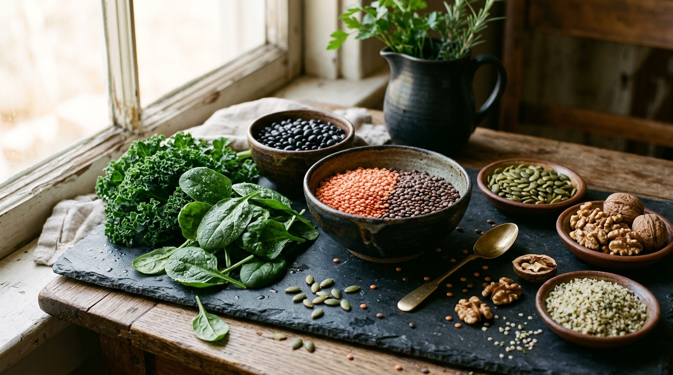
A population-based study published in Food, Nutrition and Health reports that higher dietary iron intake from natural food sources is inversely associated with the severity of abdominal aortic calcification in middle-aged and older U.S. adults. The research, utilizing data from the 2013-2014 National Health and Nutrition Examination Survey (NHANES), analyzed 2,640 participants aged 40 years and older. The findings indicate that individuals in the highest tertile of dietary iron intake had significantly lower abdominal aortic calcification scores and a reduced likelihood of severe calcification compared to those in the lowest intake group. This association was observed after adjusting for a comprehensive set of demographic, socioeconomic, lifestyle, and metabolic covariates.
Abdominal aortic calcification is a pathological condition characterized by the accumulation of mineral components within the walls of blood vessels. It is a prominent site for vascular calcification, a process that contributes to vascular sclerosis and is associated with increased cardiovascular risk. In the United States, the prevalence of abdominal aortic calcification reaches 28.8% among adults, rising to 96% in individuals over the age of 85. The study employed the Kauppila score, a semi-quantitative method derived from dual-energy X-ray absorptiometry (DXA) scans of the lateral lumbar spine, to measure the severity of calcification. Scores range from 0 to 24, with higher values indicating more extensive calcification.
The study population comprised 2,640 eligible participants after excluding individuals under 40 years of age, as well as those with missing data on dietary iron intake or calcification scores. The mean age of the participants was 57.70 years, and the cohort was composed of 52.34% females and 47.66% males. The mean abdominal aortic calcification score for the entire cohort was 1.47. The overall prevalence of any calcification was 27.79%, while the prevalence of severe calcification, defined as a score greater than 6, was 8.12%. Participants in the highest dietary iron intake tertile exhibited the lowest rate of severe calcification at 5.63%.
Dietary iron intake was assessed using two 24-hour dietary recall interviews, one conducted in person and the other by telephone. To ensure the analysis reflected intake from natural food sources, nutrients obtained from dietary supplements and medications were excluded. The researchers utilized multivariate linear and logistic regression models to evaluate the association between dietary iron intake and both the continuous calcification score and the binary outcomes of any and severe calcification. Model 3, the fully adjusted model, included covariates such as age, sex, race, education, poverty income ratio, body mass index, glycated hemoglobin, serum creatinine, and various lipid and metabolic markers.
The results demonstrated a consistent inverse association between dietary iron intake and the abdominal aortic calcification score. In the fully adjusted model, each unit increase in dietary iron intake was associated with a 0.03-unit decrease in the calcification score. More notably, participants in the highest tertile of dietary iron intake had a mean calcification score that was 0.56 units lower than those in the lowest tertile. This difference represents approximately 38% of the average calcification burden observed in the cohort, suggesting a potentially meaningful shift in disease severity at the population level.
Regarding the risk of severe abdominal aortic calcification, the study found a statistically significant inverse association. In the fully adjusted model, a single unit increase in dietary iron intake was associated with a 2% decrease in the risk of severe calcification. When comparing the highest and lowest tertiles of intake, participants in the highest group had a 60% lower risk of severe calcification. This finding was robust to sensitivity analyses, including adjustments for total dietary fiber intake, which helped control for the potential confounding effect of plant-based dietary patterns.
However, the study did not find a statistically significant association between dietary iron intake and the overall risk of any abdominal aortic calcification. The odds ratio for any calcification in the fully adjusted model was 0.98, which was not statistically significant. The authors suggest that this discrepancy may reflect endpoint heterogeneity, where the aggregation of minimal and extensive disease in the “any calcification” category dilutes specific associations. Severe calcification, representing a distinct advanced phenotype, may be more sensitive to the protective effects of adequate iron intake, particularly in the context of disease progression rather than initial nucleation.
Subgroup analyses revealed that the association between dietary iron intake and calcification scores was modified by age and alcohol consumption. The inverse association was significant among individuals aged 60 years and older, as well as among alcohol users and current smokers. Interaction tests confirmed significant interactions for age and alcohol use. Regarding severe calcification, an inverse association was noted among subjects with a history of stroke, although the interaction test was not significant, and the authors caution that this finding should be interpreted with care due to the relatively small sample size in this subgroup.
The study is cross-sectional in design, which limits the ability to infer causal relationships or temporal sequences between dietary iron intake and the development of calcification. Reverse causation cannot be ruled out, and longitudinal studies are needed to determine whether dietary iron intake influences the trajectory of calcification over time. Additionally, dietary iron intake was assessed using 24-hour recalls, which may not accurately reflect long-term habitual intake due to day-to-day variation and recall bias. The study also could not differentiate between heme and non-heme iron sources, a limitation given that these forms may have different biological effects on cardiovascular health.
The authors propose that the observed benefits may be driven by the predominant non-heme iron fraction, which constitutes the majority of total iron intake in Western diets. Within physiological limits, higher dietary iron intake likely reflects improved iron sufficiency, supporting antioxidant defenses and mitochondrial function. Iron is a crucial cofactor for enzymes involved in regulating oxidative stress, such as superoxide dismutase and catalase. By maintaining redox balance, adequate iron intake may mitigate oxidative damage to vascular cells, thereby slowing the progression of calcification. This mechanism is distinct from the risks associated with pathological iron overload, which can promote oxidative stress and vascular damage through the Fenton reaction.
The findings suggest that clinicians should consider dietary iron intake in patients at risk for abdominal aortic calcification. While the study provides evidence of an association, it does not establish a causal link or provide specific clinical recommendations for supplementation. The wide confidence intervals for some estimates, particularly in the highest intake tertile, indicate limited statistical precision, likely due to the smaller number of participants in that group. Further research with larger cohorts and longitudinal designs is necessary to verify these findings and clarify the optimal range of dietary iron intake for vascular health.
This study is the first to investigate the relationship between dietary iron intake and abdominal aortic calcification, filling a gap in the literature where the association had remained largely unexamined. Previous research has highlighted the effects of dietary iron on other cardiovascular outcomes, such as lipid metabolism and stroke risk, but the specific impact on aortic calcification had not been previously quantified in a large, nationally representative sample. The use of NHANES data, with its complex sampling design and extensive covariate adjustments, enhances the credibility of the findings and their applicability to the general U.S. adult population.
Despite the strengths of the study, including its large sample size and rigorous statistical methods, several limitations remain. The cross-sectional nature of the data precludes causal inference, and the single 24-hour dietary recall may introduce misclassification of usual dietary exposure. The study was limited to adults aged 40 and older, as DXA-based calcification measurements were not performed in younger participants, limiting generalizability to the broader adult population. Additionally, the Kauppila score does not distinguish between intimal and medial calcification, which may have different pathophysiological implications. Future studies incorporating repeated imaging and specific biomarkers of iron status will be essential to confirm the role of dietary iron in modulating the progression of vascular calcification.
Subject of Research: Association between dietary iron intake and abdominal aortic calcification severity in US adults
Article Title: Association between dietary iron intake from natural food sources and the likelihood of abdominal aortic calcification in US adults: a population-based study
Article References: Liang, D., Liu, C., & Zhang, X. (2026). Association between dietary iron intake from natural food sources and the likelihood of abdominal aortic calcification in US adults: a population-based study. Food, Nutrition and Health, 3(1), Article 14. https://doi.org/10.1007/s44403-026-00060-5
Image Credits: AI Generated
DOI: 10.1007/s44403-026-00060-5
Keywords: dietary iron, abdominal aortic calcification, NHANES, vascular health, cardiovascular risk, population-based study, calcification score, nutritional epidemiology, Association, between, dietary, iron
News Source: Daisy Hatcher. (November 8, 2026). Higher Dietary Iron Linked to Lower Severe Aortic Calcification. Scienmag.