A newly published study in wearable biosensing demonstrates that combining advanced mathematical features from electrocardiography and respiration signals can classify anxiety-inducing versus soothing video stimuli within a recorded dataset, but faces substantial hurdles when generalizing to unseen individuals. Evaluating cardiorespiratory responses across eighteen participants, researchers investigated whether nonlinear and nonstationary features derived from the Hilbert transform and discrete wavelet transform could distinguish between affective conditions. Under a participant-independent nested leave-one-subject-out cross-validation protocol, the combined cardiorespiratory model achieved a balanced accuracy of 0.532 and an area under the curve of 0.543 on the primary cohort, performing near chance level. In contrast, when the identical pipeline was applied to the independent WESAD stress-versus-baseline benchmark across fifteen participants, it achieved a balanced accuracy of 0.862 and an area under the curve of 0.935. Meanwhile, an exploratory within-dataset analysis using window-level splitting reached 0.968 accuracy, highlighting how cross-subject generalization remains a pivotal challenge in physiological classification.
The clinical impetus for this work stems from the immense burden of anxiety disorders, which affect approximately one in three American adults and increased by 25.6 percent globally during the COVID-19 pandemic. Current diagnostic methods rely almost entirely on retrospective self-reports, subjective psychological questionnaires, and infrequent clinical consultations, none of which enable continuous real-time monitoring. Because anxiety triggers clear physiological changes mediated by the autonomic nervous system—including fluctuations in cardiac rhythm, heart palpitations, perspiration, and altered breathing patterns—wearable biosensors present a compelling opportunity for objective assessment. Prior studies have demonstrated that electrocardiogram metrics like heart rate variability reflect autonomic dysregulation, while respiration signals capture breathing dynamics linked to emotional states. However, the study authors observed that existing investigations rarely integrate both modalities simultaneously. Furthermore, conventional analytical approaches frequently depend on linear descriptors, potentially missing the complex nonlinear and nonstationary dynamics embedded within continuous cardiorespiratory signals under acute psychological stimulation.
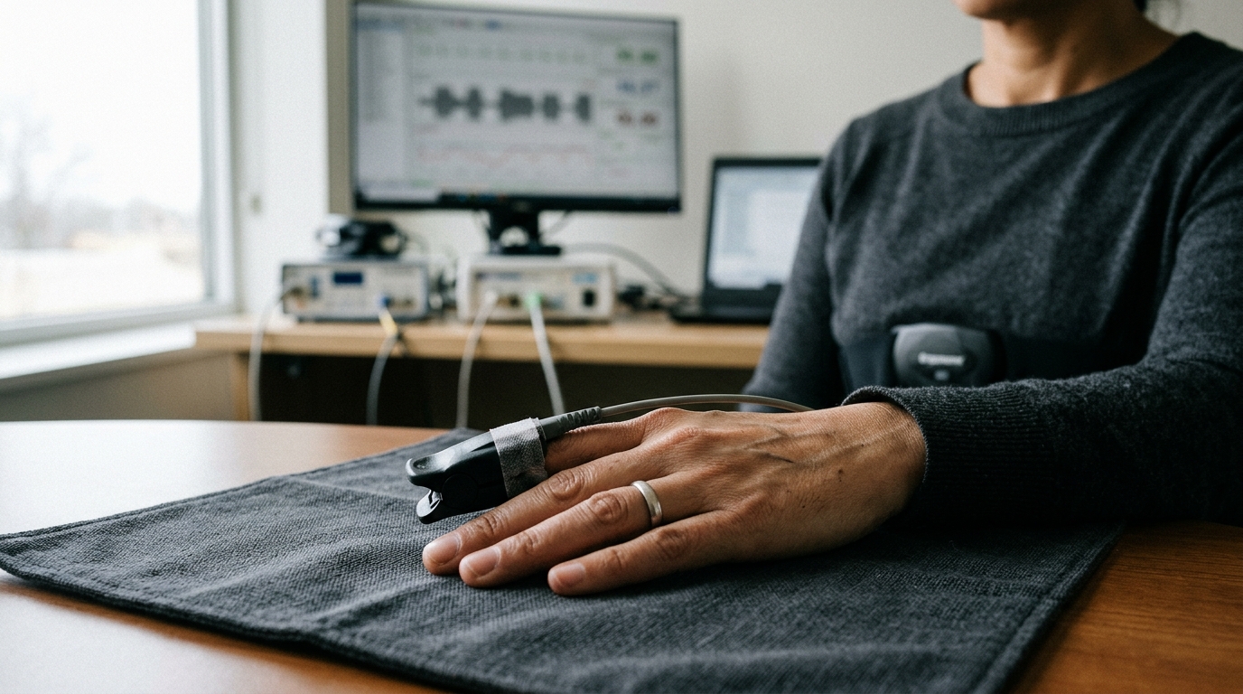
To capture these physiological responses, the research team analyzed recordings from nineteen volunteers aged between 18 and 56 years, encompassing fourteen males and five females. After excluding one participant due to excessive data artifacts, the final analyzed cohort comprised eighteen individuals. While seated comfortably, participants were fitted with wearable BIOPAC monitoring hardware that recorded electrocardiogram and respiration signals concurrently at a sampling frequency of 500 hertz. The cardiac monitor was placed on the subjects’ wrists, while a TSD201 respiratory belt secured around the chest captured thoracic movement. Volunteers watched a sequence of eight video clips in fixed alternating order. Four clips contained anxiety-inducing scenes depicting natural disasters and vehicular crashes, designed to evoke anxiety, while the remaining four clips presented calming footage, such as soothing puppy videos, not expected to induce anxiety. The first video was utilized exclusively to verify sensor attachment and signal quality, meaning data from this initial calming video were excluded from analysis, leaving seven video clips for model evaluation.
Signal preprocessing filtered and segmented the raw physiological data to prepare them for mathematical feature extraction. Electrocardiography data were filtered using an 8 to 20 hertz band-pass filter to isolate QRS energy, followed by signal squaring to enhance R-peak detection. Simultaneously, respiration signals were processed with a band-pass filter spanning 0.04 to 0.4 hertz. Signals were then divided into 60-second windows, an ultra-short-term windowing duration selected because longer recording intervals exceeded the length of most video clips. In the exploratory analysis, windows were created using a one-second sliding step and subsampled to 500 windows per participant, with each window labeled as anxiety-inducing or non-anxiety-inducing based on stimulus content. Windows at the conclusion of recording sessions were removed to avoid noise caused by participants standing up. In contrast, the participant-independent analysis generated non-overlapping 60-second windows, partitioning data strictly at the subject level before windowing occurred.
The feature engineering framework produced 58 quantitative descriptors from each 60-second window, comprising 30 conventional metrics and 28 newly proposed transform-derived features. The conventional subset included 18 heart rate variability metrics calculated from R-R intervals identified via a modified fast QRS detector, and 12 respiration rate variability metrics derived from inter-breath intervals detected using MATLAB peak-finding functions. Although standard heart rate variability analysis includes ultra-low-frequency and very-low-frequency components, these cannot be estimated reliably from 60-second epochs; a sensitivity analysis verified that excluding them did not affect participant-independent comparisons. The 28 proposed descriptors comprised 14 cardiac and 14 respiratory metrics extracted via Hilbert and wavelet transforms, marking the first time these methodologies have been jointly evaluated for classifying anxiety-inducing stimuli. The authors highlighted that these descriptors serve as empirical mathematical representations of signal structure rather than clinically validated physiological biomarkers.
The Hilbert transform extracted instantaneous amplitude, phase, and frequency components from each filtered biosignal. By constructing a complex analytic signal, the Hilbert transform yields an instantaneous amplitude envelope that captures dynamic changes in signal magnitude over time. From this envelope, four statistical features were computed: mean, standard deviation, skewness, and kurtosis. For respiration, the envelope reflects breathing depth, whereas in filtered cardiac signals, it tracks beat-to-beat modulation of QRS complex energy. The argument of the analytic signal gives the instantaneous phase, which the researchers summarized using circular statistics—specifically mean resultant length and circular phase variance—to account for the circular nature of angular data. Differentiating the temporally unwrapped phase sequence yielded instantaneous frequency, from which mean, standard deviation, skewness, and kurtosis were computed. A sensitivity analysis showed that updating phase metrics to circular statistics and unwrapping phase prior to differencing preserved high classification performance while ensuring proper mathematical formulation.
Complementing the Hilbert transform, the discrete wavelet transform provided multiresolution decomposition of cardiac and respiratory signals across varying scales and frequencies. Using the Daubechies-4 mother wavelet with a five-level decomposition and symmetric boundary extension, the algorithm separated signals into approximation and detail coefficients via iterative filtering. Four wavelet features were computed for each physiological channel: approximation coefficient mean, approximation coefficient standard deviation, wavelet entropy, and wavelet energy. The mean and standard deviation of deepest-level approximation coefficients characterized baseline signal strength and low-frequency structure. Wavelet entropy quantified structural irregularity and complexity based on the relative energy distribution across all decomposition coefficients. Wavelet energy measured the sum of squared approximation coefficients at the chosen scale, capturing the concentration of signal power. Together, these wavelet metrics captured transient fluctuations and multiscale energy dynamics, providing distinct mathematical views of cardiorespiratory responses during the experimental video conditions.
Six machine learning architectures were deployed to classify stimulus conditions: Extra Trees, Random Forest, k-Nearest Neighbors, Subspace Ensemble k-Nearest Neighbors, Support Vector Machines, and Neural Networks. In the exploratory analysis, features underwent z-score standardization computed exclusively from training data to prevent information leakage, followed by 10-fold cross-validation on an 80-20 stratified window split. Conversely, the primary participant-independent evaluation implemented an 18-fold nested leave-one-subject-out protocol where each held-out participant was completely excluded from training, imputation, feature ranking, and hyperparameter tuning. Feature selection evaluated subsets of 8, 14, 21, or 28 proposed descriptors using mutual-information ranking, with Extra Trees hyperparameters optimized through four-fold grouped cross-validation restricted to training participants. Because Extra Trees is invariant to monotonic feature scaling, standardization was omitted from the participant-independent pipeline. Model quality was evaluated using accuracy, specificity, sensitivity, precision, F1-score, and the Matthews correlation coefficient.
Experimental results revealed dramatic differences between validation frameworks. Under the exploratory within-dataset split, the Extra Trees classifier reached an accuracy of 0.968, sensitivity of 0.993, specificity of 0.883, F1-score of 0.980, and Matthews correlation coefficient of 0.908 using all 58 features. A compact subset of eight proposed transform-derived features—six cardiac and two respiratory—achieved 0.964 accuracy and an MCC of 0.894, surpassing the best sixteen-feature conventional subset at 0.943. Electrocardiogram envelope standard deviation ranked as the most important feature, followed by cardiac instantaneous frequency mean, cardiac envelope mean, cardiac envelope kurtosis, and cardiac wavelet entropy, with respiratory instantaneous frequency mean and envelope mean ranking sixth and seventh. Participant 6 demonstrated low misclassification rates during stimulus transitions. However, during the primary participant-independent evaluation on the original cohort, balanced accuracy dropped to 0.532 with an area under the curve of 0.543, showing no significant improvement over conventional features. In contrast, applying the identical pipeline to the independent WESAD stress dataset yielded a balanced accuracy of 0.862 and an area under the curve of 0.935 across fifteen unseen participants.
The authors outlined several critical limitations that define priorities for future affective computing investigations. First, the small sample size of eighteen participants restricts statistical power and limits cross-participant generalizability. Second, experimental labels reflected video stimulus content rather than clinical diagnoses or validated momentary state-anxiety ratings. Third, presenting video clips in a fixed sequential order prevented researchers from separating genuine affective responses from confounding factors like stimulus ordering, habituation, and visual fatigue. Placing their work alongside earlier studies by Desai, Ihmig, and Gazi, the authors noted that reported performance varies substantially depending on whether models are evaluated within participants or across unseen individuals. Although the WESAD evaluation demonstrated that the feature representation generalizes in an affective stress task, stress classification cannot be conflated with anxiety detection. The authors concluded that future research requires larger counterbalanced cohorts and validated psychological assessments before mathematical transform descriptors can be considered verified physiological biomarkers or translated into clinical applications.
Subject of Research: Machine learning classification of anxiety-inducing stimuli using cardiorespiratory features
Article Title: Hilbert- and wavelet-derived cardiorespiratory features for machine learning classification of anxiety-inducing stimuli
Article References: Elgendi, M., Elkhalifa, A., Stöckli, V., & Menon, C. (2026). Hilbert- and wavelet-derived cardiorespiratory features for machine learning classification of anxiety-inducing stimuli. Discover Artificial Intelligence, 6(1), Article 1816. https://doi.org/10.1007/s44163-026-02650-w
Image Credits: AI Generated
DOI: 10.1007/s44163-026-02650-w
Keywords: machine learning, electrocardiography, respiration, anxiety stimuli, Hilbert transform, wavelet transform, wearable sensors, biomedical signal processing, Hilbert-, wavelet-derived, cardiorespiratory, features
News Source: Teresa Odom. (November 7, 2026). Cardiorespiratory Features Evaluated for Machine Learning Classification of Anxiety Stimuli. Scienmag.