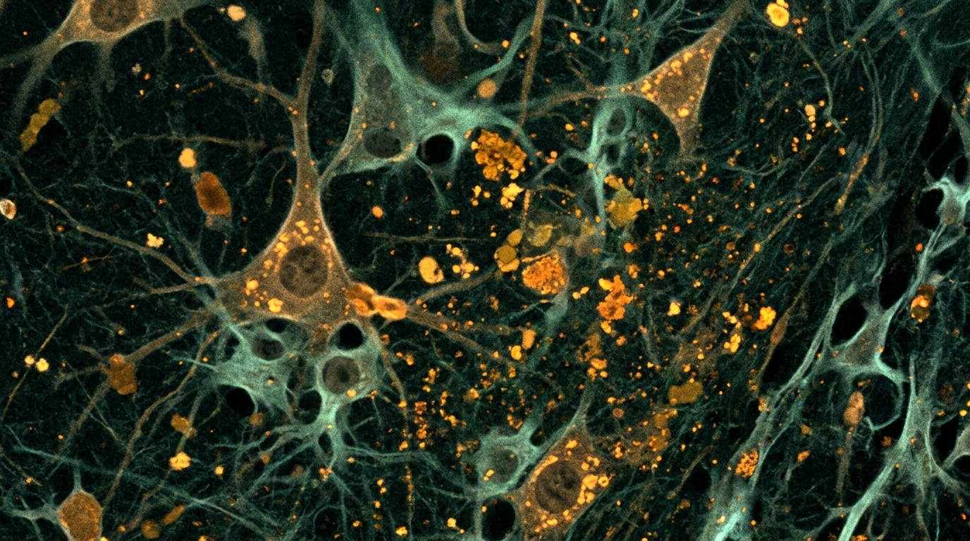
Researchers at RMIT University have identified a reproducible photophysical fingerprint of brain ageing in mice, linking specific changes in neural autofluorescence to alterations in circulating plasma lipids. The study demonstrates that as mice age, the autofluorescent material within their brains shifts toward a red-emission spectrum and exhibits shorter fluorescence lifetimes. These optical changes are not isolated to the central nervous system; they correlate significantly with the abundance of specific lipid species in the blood, suggesting a measurable connection between central brain ageing and peripheral biochemical states.
The investigation focused on the phenomenon of inflammaging, a state of chronic low-grade inflammation that characterises healthy ageing and is driven by factors such as oxidative stress and cellular senescence. In the brain, this process involves microglia, the innate immune cells responsible for maintaining tissue homeostasis. With age, microglia adopt a primed phenotype, becoming more phagocytic and overproducing cytokines in response to stimuli. The study aimed to determine whether this microglial ageing could be delineated by a quantifiable biophysical phenotype, specifically through the accumulation of autofluorescent aggregates.
Autofluorescent material, often classified under the umbrella term lipofuscin, consists of mixed aggregates of oxidised lipids, misfolded proteins, and metals. Previous work suggested that microglia in healthy ageing accumulate this material, which displays broad excitation and emission profiles. However, it remained unclear whether this accumulation reflected a structurally and spectrally distinct ageing phenotype that could be quantitatively defined. The researchers hypothesised that ageing is associated with the accumulation of this material inside microglia and that this accumulation is reflected in alterations to peripheral lipids.
To test this hypothesis, the study utilised naïve female C57BL/6 mice, comparing a young cohort aged three months to an aged cohort aged 21 to 22 months. The researchers examined the hippocampus, a region critical for memory, and the arcuate nucleus of the hypothalamus, a region associated with inflammation. Tissues were processed for quantitative real-time PCR, immunohistochemistry, and advanced optical imaging. The mice displayed expected ageing profiles, including a significant reduction in doublecortin-positive immature neurons in the hippocampus and increased microglial complexity in the arcuate nucleus.
Using confocal microscopy, the team found that all sampled microglia contained at least some autofluorescent material. In aged mice, the mean size of these aggregates increased significantly in the dentate gyrus, CA3, and arcuate nucleus regions. While young brains predominantly contained small autofluorescent particles, aged brains accumulated a higher proportion of medium, large, and extra-large aggregates. This shift in size distribution was statistically significant, indicating that microglia progressively accumulate larger autofluorescent deposits as they age.
To characterise the optical properties of these aggregates, the researchers employed photoluminescence spectroscopy and fluorescence lifetime imaging microscopy. Spectral analysis revealed that aged, large autofluorescent aggregates had a significantly lower mean intensity ratio between blue and red spectral windows compared to young, small aggregates. This indicates a relatively red-shifted emission spectrum in the aged brain. Furthermore, fluorescence lifetime imaging showed that autofluorescence in aged mice had a significantly shorter intensity-weighted average lifetime across all emission windows, including the full spectrum and specific bandpass filters.
The study then investigated whether these central optical changes were mirrored by peripheral biochemical shifts. Plasma samples from the same mice were subjected to targeted lipidomic analysis using liquid chromatography-triple quadrupole mass spectrometry. The analysis identified 22 plasma lipids that changed significantly with age. Among these, 12 lipids were significantly associated with brain autofluorescence spectra independently of age. Two lipids, sphingomyelin 32:2 (SM 32:2) and phosphatidylethanolamine 38:6 (PE 38:6), survived Bonferroni correction for multiple comparisons, demonstrating a robust statistical association with the spectral profile of brain autofluorescence.
To validate these findings, the researchers replicated the spectral and lipidomic analyses in a separate cohort of ageing mice. The validation cohort confirmed the spectral red-shift in aged brain autofluorescence. In this independent group, SM 32:2 remained significantly associated with the brain autofluorescence spectral profile, accounting for 57% of the variance in the spectral ratio. However, PE 38:6 did not show a significant relationship in the validation cohort, highlighting SM 32:2 as a more consistent peripheral marker. The consistency of the spectral shift across cohorts supports the idea that these photophysical changes are a robust feature of brain ageing.
The association between brain autofluorescence and plasma lipids suggests a bidirectional relationship between central and peripheral biochemical ageing processes. SM 32:2 is a sphingomyelin that has been implicated in inflammatory and immune responses, vascular homeostasis, and lysosomal function. In humans, SM levels are associated with memory impairment in ageing and subclinical atherosclerosis. The increase in SM 32:2 in aged mice may reflect systemic lipidomic adaptations to pathological and ageing-related inflammatory stress, potentially linking the optical signature of the brain to broader metabolic changes.
The underlying mechanisms for these changes likely involve lysosomal and autophagic decline. Ageing is associated with defective lysosomal acidification and impaired autophagic clearance, which can lead to the buildup of cross-linked, undegradable biomaterials within cells. Oxidative stress drives lipid peroxidation, contributing to the formation of autofluorescent aggregates. The shorter fluorescence lifetimes observed in aged tissue may reflect changes in the local biochemical environment, such as pH levels or ion concentrations, which influence the radiative decay of endogenous fluorophores. These factors collectively create a self-perpetuating cycle of oxidative stress and impaired degradation.
The study has several limitations that must be considered when interpreting the results. The research was conducted exclusively in female mice, and it is known that female mice exhibit higher neuroinflammation compared to males even at young ages. Additionally, plasma levels of SM 32:2 are significantly higher in female than male humans, which may limit the generalisability of this specific lipid as a biomarker to other populations. The study also relied on a relatively small sample size, and the association between lipids and autofluorescence does not establish a causal direction. Further investigation is needed to determine how these biochemical dynamics shape autofluorescence properties and to test the generalisability of these findings in male cohorts and other animal models.
Despite these limitations, the study provides a quantitative basis for tracking early physiological changes associated with neurodegenerative risk. By integrating autofluorescence spectroscopy and lifetime imaging with plasma lipidomics, the researchers have established a biophotonic phenotype of ageing. This approach offers a new dimension to ageing biology, bridging molecular pathology with optical physics. The identification of specific spectral and lifetime signatures, coupled with peripheral lipid markers, opens the path toward future non-invasive optical biomarkers for monitoring brain ageing and preclinical neurodegeneration before structural or cognitive decline becomes detectable.
Beyond structural autofluorescence, the researchers confirmed the presence of distinct molecular shifts in hippocampal tissue using quantitative real-time PCR. Aged mice exhibited significant upregulation of inflammation-related genes, including Itgam, Tlr4, Aif1, and MhcII, pointing to heightened immune activity. Concurrently, genes associated with Alzheimer’s disease demonstrated divergent regulation: expression of App and the endolysosomal trafficking regulator Sorl1 declined, whereas Mapt and Abca7 expression increased. Lipid metabolism genes Hexb, Ch25h, and Lpl were also significantly elevated in older animals. These transcriptional changes accompanied marked differences in hippocampal neurogenesis. While the total number of mature, NeuN-positive neurons remained unchanged between young and aged mice, doublecortin-positive immature neurons declined sharply in the dentate gyrus across all developmental stages, including proliferative, intermediate, and post-mitotic cells.
Morphological evaluations using Sholl analysis further revealed regional variations in microglial remodeling. Although microglial cell counts rose in the dentate gyrus, their branching numbers, total process lengths, and overall complexity were unaltered by age. Similarly, microglia in the CA1 and CA3 regions showed no substantial differences in branch count or complexity. In contrast, arcuate nucleus microglia displayed both elevated cell numbers and greater structural complexity, particularly showing increased branching within a radius of one to ten micrometres from the cell soma. Autofluorescence volume also correlated differently with microglial morphology depending on brain anatomy. While no relationship emerged between branching complexity and autofluorescent volume in hippocampal regions, microglial branch points in the arcuate nucleus exhibited a moderate, statistically significant positive correlation with the mean volume of internal autofluorescent aggregates.
To resolve the complex emission signals, the researchers computed the second derivative of the photoluminescence spectra, identifying eighteen individual emission peaks between 507 and 731 nanometres. Nine prominent peaks appeared consistently across more than ninety percent of examined animals, situated at 507, 546, 557, 574, 589, 608, 628, 649, and 731 nanometres. The relative magnitude of these derivatives differed significantly between age groups; for instance, the peak at 507 nanometres was substantially more pronounced in young, small aggregates than in aged deposits, and the 546 and 731 nanometre peaks similarly showed reduced prominence in aged tissue. Time-resolved measurements supplied further physical specificity, establishing that intensity-weighted autofluorescence lifetimes across the full 500 to 750 nanometre detection window averaged 2.43 nanoseconds in young tissue compared to 2.09 nanoseconds in aged tissue.
The secondary validation cohort, comprising six young and seven aged female mice assessed six months later, reinforced these spectral patterns while extending exploratory plasma observations. Although one aged animal was identified post-mortem with a brain tumour, re-evaluating the dataset with or without that outlier confirmed the consistency of the spectral red shift. Beyond the robust association observed for sphingomyelin 32:2, exploratory regressions in this cohort indicated that three additional plasma lipids tracked the brain optical shift: lower levels of acylcarnitine 16:0 alongside higher concentrations of phosphatidylethanolamine 36:4 and phosphatidylethanolamine 38:4 accompanied more red-shifted cerebral autofluorescence.
Subject of Research: Establishing a Biophotonic Signature of Brain Ageing
Article Title: Establishing a Biophotonic Signature of Brain Ageing
Article References: Slayo, M., Vocale, L., Xavier, S., Singhaarachchi, P., Quinn, K. M., del Rosal, B., & Spencer, S. J. (2026). Establishing a Biophotonic Signature of Brain Ageing. Aging Cell, 25(11), Article e70790. https://doi.org/10.1111/acel.70790
Image Credits: AI Generated
DOI: 10.1111/acel.70790
Keywords: Establishing, Biophotonic, Signature, Brain, Ageing, scientific research
News Source: Cassandra Pierce. (November 7, 2026). Mouse Study Links Brain Autofluorescence Shifts to Plasma Lipids. Scienmag.