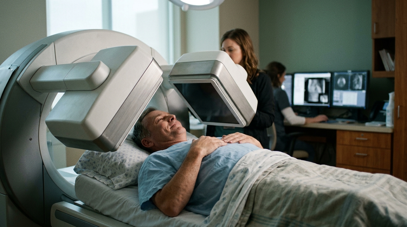
A multicenter study evaluating diagnostic imaging for transthyretin cardiac amyloidosis has demonstrated that combining routine clinical variables with planar scintigraphy scans through a multimodal deep learning framework improves the accuracy of predicting expert-adjudicated Perugini uptake categories. Technetium-99m pyrophosphate planar scintigraphy is widely utilized to assess suspected transthyretin cardiac amyloidosis, but interpreting equivocal Perugini Grade 1 cardiac uptake frequently presents a challenge in clinical workflows due to low signal-to-noise ratios and overlapping blood-pool activity. To address this ambiguity, researchers developed and evaluated a deep learning system specifically designed to predict single-photon emission computed tomography and computed tomography adjudicated categories directly from planar images and pre-procedural clinical data.
The investigation utilized clinical records and imaging data from 438 patients registered in the Taiwan Multicenter TTCA-PYP Registry. The primary target was the classification of cases into three expert SPECT/CT-adjudicated Perugini groups: Grade 0, Grade 1, and Grades 2 to 3. The investigators specifically noted that patient genetic testing status and study design characteristics were intentionally excluded from the input features, focusing the model exclusively on readily accessible pre-procedural parameters alongside scintigraphic regions of interest.
To process these diverse data streams, the framework incorporated specialized encoders joined by an attention mechanism. Physician-verified planar cardiac regions of interest measuring 32 by 128 pixels were analyzed using a truncated Wide-ResNet50-2 convolutional neural network. Simultaneously, 23 routine clinical variables were represented using a frozen TabPFN tabular foundation model embedding. The image representations and clinical feature embeddings were aligned during training using a contrastive loss inspired by CLIP architecture and subsequently integrated through cross-attention to predict the final Perugini uptake classifications.
Model performance was evaluated using ten repetitions of a hospital- and grade-stratified assessment set alongside ten-fold cross-validation, with statistical intervals calculated using a Nadeau-Bengio correction for repeated resampling. Decision curve analysis was also conducted to gauge clinical utility. The multimodal approach achieved a higher overall classification accuracy of 0.826 compared to 0.751 achieved by an imaging-only baseline, representing a paired difference of 0.075 with a 95 percent confidence interval from 0.034 to 0.117. Meanwhile, the macro-averaged area under the receiver operating characteristic curve remained similar between the two approaches, recorded at 0.925 for the multimodal system versus 0.917 for the imaging-only model.
The evaluation focused particular attention on intermediate Grade 1 scans, where diagnostic interpretation is historically most equivocal. In this subset, the multimodal model increased classification precision from 0.536 under the image-only model to 0.694, yielding a precision difference of 0.159 with a 95 percent confidence interval between 0.056 and 0.261. However, this gain was accompanied by a slight drop in Grade 1 recall, which decreased from 0.704 to 0.683, representing a difference of minus 0.022 with a 95 percent confidence interval ranging from minus 0.120 to 0.077. The researchers observed that shifts in F1-scores and precision-recall area under the curve metrics showed less certainty.
Generalization across medical centers exposed notable limitations during leave-one-center-out testing across the two participating hospitals containing all diagnostic grades, Far Eastern Memorial Hospital and National Taiwan University Hospital. While held-out overall accuracy reached 0.704 for FEMH and 0.889 for NTUH, sensitivity varied considerably: Grade 1 recall dropped to 0.312 when evaluating FEMH as the held-out center, while Grade 0 recall fell to 0.071 when NTUH was held out. The authors concluded that while the multimodal network may serve as an adjunct to standardize imaging-based Perugini categorization from planar Tc-99m PYP examinations, it does not establish an etiologic diagnosis of transthyretin cardiac amyloidosis. Grade 1 predictions remain indeterminate classifications that continue to require monoclonal protein testing and further tomographic or clinical evaluation, underscoring the necessity of broader independent validation before clinical implementation.
Subject of Research: Multimodal deep learning for Perugini category prediction in Tc-99m PYP planar scintigraphy
Article Title: Integration of clinical parameters and deep learning based ROI enhancement in Tc-99m PYP scintigraphy for predicting SPECT/CT-adjudicated Perugini categories in suspected transthyretin cardiac amyloidosis
Article References: Ou, D.-X., Huang, C.-H., Wang, S.-Y., Lin, Y.-H., Chang, W.-T., Yu, W.-C., Shih, J.-Y., Chang, K.-C., Ko, C.-L., Cheng, M.-F., Yen, Y.-A., Hu, L.-H., Kao, C.-H., Tsai, S.-Y., Lai, C.-H., Hung, C.-L., Wang, W.-H., Chen, C.-M., Wu, Y.-W., & on behalf of the TTCA-PYP investigators (2026). Integration of clinical parameters and deep learning based ROI enhancement in Tc-99m PYP scintigraphy for predicting SPECT/CT-adjudicated Perugini categories in suspected transthyretin cardiac amyloidosis. BMC Medical Imaging. https://doi.org/10.1186/s12880-026-03036-1
Image Credits: AI Generated
DOI: 10.1186/s12880-026-03036-1
Keywords: transthyretin cardiac amyloidosis, Tc-99m PYP scintigraphy, Perugini classification, deep learning, SPECT/CT, multimodal AI, Wide-ResNet50-2, TabPFN, Integration, clinical, parameters, deep
News Source: Ophelia Keating. (November 7, 2026). Multimodal AI Model Improves Cardiac Amyloidosis Classification from Planar Scintigraphy. Scienmag.