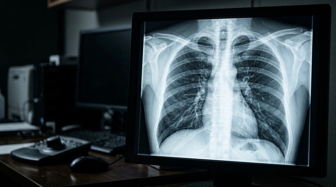
Deep learning algorithms deployed for automated tuberculosis detection experience substantial declines in diagnostic sensitivity and probability calibration when evaluated on chest radiographs subjected to common synthetic image quality defects, according to an experimental investigation published in the Egyptian Journal of Radiology and Nuclear Medicine. Researchers Nitin Wankhade, Sagar Joshi, Ashish Manwatkar, Dnyaneshwar Kudande, Nitin Dhawas, and Dheeraj Patil conducted a failure-aware evaluation to measure how performance drops when convolutional neural networks process corrupted clinical imagery rather than pristine, curated benchmark datasets. While artificial intelligence systems have consistently demonstrated high diagnostic performance in controlled academic settings, images gathered during routine clinical screening in resource-limited regions frequently suffer from physical, optical, and digital transmission artifacts. By systematically corrupting held-out test data across multiple architectures and severity tiers, the authors documented that image deterioration disproportionately increases false-negative outcomes and diminishes the reliability of network confidence values.
The study addressed persistent real-world operational challenges identified by global health authorities, where tuberculosis continues to present a major public health challenge responsible for roughly ten million new infections and over one million deaths every year. Because standard screening regimens rely on cost-effective chest radiography to identify early radiological signs, automated computational tools are increasingly considered for screening programs in community settings, mobile radiography vans, and remote clinical outposts facing severe shortages of trained radiological specialists. Despite encouraging benchmark scores where areas under the receiver operating characteristic curve frequently exceed 0.85, an operational gap exists between academic evaluations and clinical deployment environments. Routine acquisition workflows frequently introduce linear motion blur caused by patient movement during radiographic exposure, quantum noise and reduced contrast stemming from suboptimal exposure settings or absent scatter grids, resolution downsampling during digital transmission, and block compression artifacts from digital storage archiving pipelines.
To evaluate these operational failure modes, the researchers assembled a pooled benchmark cohort comprising 800 posterior-anterior chest radiographs drawn from two widely recognized public repositories: the Montgomery County X-ray Set and the Shenzhen Hospital X-ray Set. The Montgomery collection provided 138 radiographs originating from the Department of Health and Human Services in Montgomery County, Maryland, encompassing 58 tuberculosis-positive cases alongside radiologist reading notes and 80 normal examinations. The Shenzhen collection, derived from Shenzhen No. 3 People’s Hospital in China, contributed 662 radiographs comprising 336 confirmed tuberculosis-positive images and 326 negative cases. Across the unified 800-patient pool, the disease prevalence stood at 49.25 percent, representing exactly 394 confirmed tuberculosis cases and 406 non-tuberculosis examinations, with each individual patient contributing exactly one radiograph to eliminate duplicate anatomical sampling.
The experimental methodology implemented a conventional, non-repeated stratified fivefold cross-validation scheme to guard against data leakage across training, validation, and testing partitions. Within each partition, images were allocated along an image-level and patient-level split of 70 percent for training, 10 percent for internal validation, and 20 percent for testing, representing roughly 560 training images, 80 validation images, and 160 held-out test cases per fold. Each held-out test fold preserved balanced proportions consisting of 78 to 79 tuberculosis-positive images and 81 to 82 negative images, drawn consistently from both source repositories. The research team evaluated three convolutional neural network architectures representing distinct structural paradigms: ResNet-50 incorporating residual skip connections, DenseNet-121 utilizing dense feature connectivity, and MobileNetV2 employing depth-wise separable convolutions to minimize computational footprints.
Model training executed on a single graphics processing unit utilizing ImageNet-pretrained baseline weights fine-tuned end-to-end without frozen layers, optimizing binary cross-entropy loss through the Adam optimizer under an initial learning rate of 0.0001 and a batch size of 16. To preserve experimental control and isolate the specific effects of synthetic degradation, the preprocessing pipeline deliberately avoided supplementary denoising, edge sharpening, or class-weighting adjustments. Grayscale images were normalized to zero-to-one floating-point intervals through per-image min-max scaling, resized to a standardized 224 by 224 pixel input resolution via bilinear interpolation, and expanded to three identical channels. Decision thresholds were fixed independently for each cross-validation fold by maximizing Youden’s J statistic exclusively on internal validation subsets, ensuring that subsequent changes in threshold-dependent clinical performance reflected image corruption rather than post-hoc threshold manipulation.
The evaluation framework modeled five distinct synthetic degradation types across mild, moderate, and severe degradation levels, applied exclusively to held-out test sets while training and validation data remained completely pristine. The perturbation battery included additive Gaussian noise to simulate quantum variations and electronic sensor readout noise, linear motion blur kernels to replicate patient movement during exposure, lossy Joint Photographic Experts Group compression to reproduce archiving quantization, spatial downsampling down to 112 by 112 pixels to simulate transmission bottlenecks, and nonlinear gamma contrast reduction to approximate scattered radiation effects. The degradation levels were applied independently rather than cumulatively, enabling isolated robustness profiling across forty-five distinct model, perturbation, and severity permutations while tracking discrimination, positive predictive values, negative predictive values, and expected calibration errors.
On pristine test data, all three deep learning architectures established strong baseline discrimination, achieving fold-averaged area under the receiver operating characteristic curve values ranging from 0.89 to 0.93 alongside expected calibration errors between 0.038 and 0.051. DenseNet-121 demonstrated the strongest pristine discrimination with an area under the curve of 0.93, while MobileNetV2 registered slightly lower baseline accuracy. However, the introduction of synthetic degradations degraded diagnostic capability across every evaluated model. Linear motion blur produced the most pronounced operational damage, reducing sensitivity by an average relative drop exceeding 15 percent across all networks and causing relative sensitivity drops of up to 21.3 percent in DenseNet-121. Spatial downsampling represented the second largest source of sensitivity loss, generating an average relative drop of 12.9 percent in DenseNet-121, whereas Gaussian noise and compression artifacts produced comparatively smaller relative reductions.
Further quantitative examination demonstrated asymmetric vulnerability between true-positive identification and negative-case screening across degraded testing environments. While positive predictive value suffered severe declines ranging between 13.6 percent and 20.9 percent across the evaluated architectures, negative predictive value dropped by only 5.6 percent to 6.6 percent under moderate degradation. Across the aggregated moderate degradation conditions, receiver operating characteristic values fell by 0.06 to 0.08, while expected calibration error increased by a factor of 1.7 to 2.7, climbing to ranges between 0.086 and 0.104 across architectures. Error distribution profiling revealed that the rate of false negatives nearly tripled under severe motion blur in DenseNet-121, driven primarily by the obscuration of subtle, low-contrast parenchymal infiltrates and cavitary lesions that became visually indistinguishable from surrounding lung tissue once smeared by motion convolution.
Comparative structural analysis revealed a trade-off between clean benchmark diagnostic accuracy and relative resilience under synthetic imaging degradation. Although DenseNet-121 achieved the highest pristine performance, it exhibited the steepest sensitivity loss under motion blur and a 2.7-fold increase in calibration error. Conversely, MobileNetV2 recorded the smallest mean relative sensitivity drop of 10.6 percent across moderate degradations, though this apparent stability reflected a lower clean baseline rather than superior absolute detection under stress. The researchers hypothesized that spatial blurring degrades performance by exceeding the effective receptive field of early convolutional feature extractors and distorting learned penultimate-layer representations, as demonstrated by measurable Euclidean feature vector shifts observed in DenseNet-121 under moderate motion blur.
The study authors highlighted several methodological limitations, noting that synthetic perturbations cannot fully capture the multifaceted, coupled artifacts and hardware-specific point-spread variations seen in physical clinical environments. Furthermore, because image corruptions were applied after initial normalization and resizing to 224 by 224 pixels rather than at native detector resolutions, the experimental noise parameters describe network-space perturbations rather than physical radiation measurements. To bridge the gap between academic benchmarks and clinical deployment, the investigators proposed phased future research involving prospective validation across five thousand naturalistic mobile radiography scans, the exploration of multiscale and vision transformer architectures to compress sensitivity gaps below five percentage points, and the implementation of automated image quality triage pipelines before automated inference.
Subject of Research: Deep learning robustness and calibration for automated tuberculosis detection under synthetic chest radiograph degradation
Article Title: Failure-aware robustness evaluation of deep learning models for tuberculosis detection under synthetic chest X-ray degradation
Article References: Wankhade, N., Joshi, S., Manwatkar, A., Kudande, D., Dhawas, N., & Patil, D. (2026). Failure-aware robustness evaluation of deep learning models for tuberculosis detection under synthetic chest X-ray degradation. Egyptian Journal of Radiology and Nuclear Medicine, 57(1), Article 238. https://doi.org/10.1186/s43055-026-01902-0
Image Credits: AI Generated
DOI: 10.1186/s43055-026-01902-0
Keywords: tuberculosis detection, chest radiography, deep learning, convolutional neural networks, image degradation, model robustness, expected calibration error, diagnostic sensitivity, medical imaging, artificial intelligence, Failure-aware, robustness
News Source: Ophelia Keating. (November 7, 2026). Degraded Chest Radiographs Reduce Diagnostic Sensitivity in Deep Learning Tuberculosis Detection Models. Scienmag.