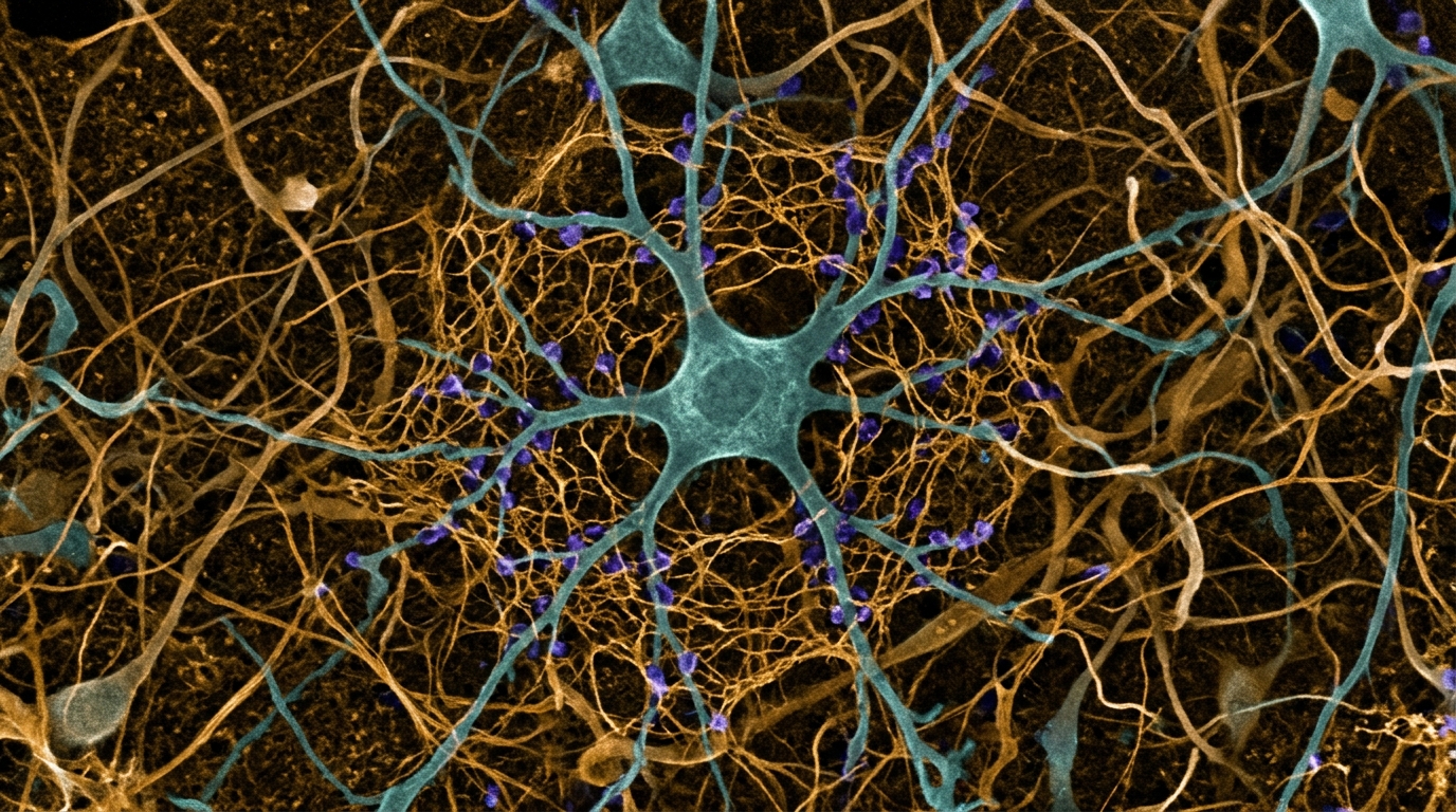
In a newly published hypothesis and theory review in Frontiers in Molecular Neuroscience, researchers Wu Yisen, Chen Xiao, and Lu Song propose that perineuronal nets in the spinal dorsal horn act as an active molecular brake on maladaptive neurotrophin signaling during chronic neuropathic pain. Drawing on established neurobiological literature and mechanistic biochemical models, the authors reframe these dense extracellular matrix structures from passive physical scaffolds into dynamic regulators of postsynaptic inhibitory efficacy. Neuropathic pain, defined as pain arising from a lesion or disease of the somatosensory nervous system, frequently becomes autonomous of ongoing peripheral injury once central sensitization is established in the central nervous system. A central driver of this autonomous hyperexcitability is spinal disinhibition, where the loss of inhibitory control permits innocuous tactile inputs from low-threshold mechanoreceptors to engage pain transmission circuits. The authors synthesize published molecular and circuit studies to formulate a testable theoretical model in which intact perineuronal nets constitutively suppress pathological signaling cascades within nociceptive projection neurons, whereas injury-induced net degradation unleashes molecular disinhibition.
The conceptual framework centers on a two-cell microcircuit in the superficial dorsal horn, comprising a presynaptic parvalbumin-positive inhibitory interneuron and a postsynaptic lamina I projection neuron. Under physiological conditions, parvalbumin-positive interneurons function as circuit-level gatekeepers that prevent tactile inputs carried by low-threshold mechanoreceptors from reaching projection pathways. In their theoretical formulation, the authors distinguish this circuit gatekeeping role from a molecular brake mechanism, which they assign to protein tyrosine phosphatase sigma and its interaction with perineuronal nets surrounding the projection neuron. Perineuronal nets consist of a hyaluronan backbone cross-linked with chondroitin sulfate proteoglycans such as aggrecan, brevican, neurocan, and versican, alongside link proteins and tenascin-R. The highly sulfated glycosaminoglycan chains of these proteoglycans display high affinity for receptor protein tyrosine phosphatase sigma, an enzyme that clusters at cell surfaces upon binding specific proteoglycan motifs. The authors posit that this extracellular matrix lattice positions the phosphatase to constitutively dephosphorylate nearby receptor tyrosine kinases, specifically dampening selected downstream effectors of tropomyosin receptor kinase B.
According to the proposed model, intact perineuronal nets exert their regulatory influence through two complementary biophysical and biochemical processes. Physically, the dense extracellular matrix forms a diffusion barrier that restricts the mobility and local concentration of brain-derived neurotrophic factor, thereby limiting the duration and intensity of neurotrophin receptor engagement. Enzymatically, chondroitin sulfate proteoglycan binding to protein tyrosine phosphatase sigma sustains tonic phosphatase activity directed toward membrane-proximal receptor substrates. The authors specifically hypothesize that this phosphatase activity selectively dephosphorylates tyrosine residue 816 on tropomyosin receptor kinase B, which serves as the docking site for phospholipase C-gamma-1, while sparing phosphorylation at tyrosine residue 515, which mediates trophic signaling through the extracellular signal-regulated kinase and Akt pathways. Under baseline conditions, this putative selective dephosphorylation prevents overactivation of intracellular calcium mobilization pathways while maintaining neurotrophic support necessary for cellular survival. The authors explicitly identify this residue-selective dephosphorylation as an untested hypothesis, emphasizing that direct biochemical proof in nerve injury models has not yet been demonstrated.
Following peripheral nerve injury, the proposed molecular brake fails as spinal perineuronal nets undergo structural degradation mediated by reactive neuroglia. Injured primary sensory afferents and reactive spinal microglia release extracellular proteases, including matrix metalloproteinase-9 and cathepsin S, which cleave proteoglycan core proteins, while microglia also actively engulf and remove net fragments through phagocytosis. The authors detail how this degradation attenuates the physical barrier against ligand diffusion and disrupts proteoglycan-mediated engagement with protein tyrosine phosphatase sigma. Relieved of tonic phosphatase restraint, tropomyosin receptor kinase B receptors exhibit amplified or prolonged phosphorylation at tyrosine residue 816 upon exposure to basal or microglia-derived brain-derived neurotrophic factor. The authors clarify that this brake release modulates ligand-driven signaling rather than causing ligand-independent receptor activation, as neurotrophin binding remains requisite for receptor dimerization. Once unchecked, the activated phospholipase C-gamma-1 cascade hydrolyzes phosphatidylinositol 4,5-bisphosphate into inositol trisphosphate and diacylglycerol, triggering intracellular calcium release and establishing a signaling platform that initiates postsynaptic transporter disruption.
The downstream consequence of this amplified signaling cascade converges on the potassium-chloride cotransporter 2, designated KCC2, which maintains low intracellular chloride concentrations in mature neurons. The theoretical model posits that elevated intracellular calcium activates calpain proteases, which recognize specific sequences in the KCC2 cytosolic domain and execute proteolytic cleavage. Detached from the cell membrane, cleaved transporters undergo degradation, diminishing neuronal chloride extrusion capacity. The resulting intracellular chloride accumulation alters the reversal potential of GABA type A and glycine receptor-mediated currents in a depolarizing direction. Because depolarization reduces the hyperpolarizing driving force, postsynaptic inhibition is weakened or converted under defined physiological conditions into an excitatory influence. In their evidence classification, the authors emphasize that while calpain-dependent KCC2 cleavage has been directly demonstrated through NMDA receptor and activity-dependent pathways, the specific link connecting neurotrophin-dependent phospholipase C-gamma-1 signaling directly to calpain activation in dorsal horn projection neurons remains an unverified hypothesis.
At the circuit level, the proposed postsynaptic loss of KCC2 converges with presynaptic dysfunction occurring within parvalbumin-positive interneurons, uniting both cellular compartments around calcium dysregulation. Following peripheral nerve injury, parvalbumin protein levels decline in dorsal horn interneurons, which impairs cytosolic calcium buffering and recruits small-conductance calcium-activated potassium channels. This presynaptic channel activation converts continuous tonic firing into adapting discharge patterns, decreasing sustained release of inhibitory neurotransmitters. Simultaneously, the postsynaptic projection neuron experiences diminished inhibitory sensitivity due to chloride extrusion breakdown driven by matrix degradation and neurotrophin signaling. The authors conceptualize this dual failure as a shared breakdown of calcium homeostasis across the gatekeeper microcircuit, wherein presynaptic firing output is muted while postsynaptic receptive efficacy is degraded. They underline that because these presynaptic and postsynaptic events have not been demonstrated concurrently within a single validated cellular preparation, their integration constitutes a circuit-level hypothesis requiring multi-cell electrophysiological validation.
To contextualize the temporal progression of these events, the review reconstructs a dynamic chronology synthesized from diverse rodent injury models. In the acute window spanning the first 24 hours post-injury, perineuronal net structure remains grossly intact while microglial activation and early protease induction commence. During the early subacute period between 24 and 72 hours, matrix metalloproteinase-9 activity reaches peak levels, initial proteoglycan cleavage begins, and early tactile allodynia emerges alongside complement deposition. By the late subacute phase from 3 to 7 days, microglial phagocytosis of extracellular matrix fragments accelerates, leading to pronounced net loss that coincides with peak microglial neurotrophin release. The early chronic phase, spanning 7 to 14 days, corresponds to maximal calpain-mediated transporter cleavage and sustained behavioral allodynia, which subsequently transitions after two weeks into chronic maintenance reinforced by transcriptional repression. The authors stress that this timeline represents a conceptual compilation derived across varied experimental paradigms rather than a continuous sequence observed longitudinally within a single standardized animal cohort.
In evaluating translational strategies, the authors examine pharmacological tools that intersect with the proposed pathway, drawing strict distinctions between experimental probes and viable therapeutics. The intracellular sigma peptide, a cell-permeable agent designed to disrupt protein tyrosine phosphatase sigma wedge-domain signaling, has demonstrated efficacy in promoting axonal sprouting and functional recovery in spinal cord injury. However, within the logic of the proposed pain model, inhibiting this phosphatase would remove the molecular brake on receptor phosphorylation, potentially exacerbating phospholipase activation and accelerating transporter degradation. Consequently, the authors treat the intracellular sigma peptide strictly as an experimental tool to test pathway causality rather than as a candidate analgesic. Conversely, direct enhancement of chloride extrusion represents a translationally coherent approach, exemplified by small-molecule KCC2 enhancers such as CLP290 and kenpaullone, which restore chloride transport and reduce pain behaviors in preclinical rodent assays without provoking sedation or motor impairment.
The review delineates several critical limitations, alternative explanations, and evidence gaps that must temper immediate conclusions. Most prominent is the classical neurotrophin-centric model, which demonstrates that microglial neurotrophin release is sufficient to trigger disinhibition independently of matrix remodeling, meaning the relative necessity of net degradation remains unsettled. The authors formulate falsification criteria to test whether matrix loss alone is sufficient to induce disinhibition, whether neurotrophin binding remains strictly necessary, or whether synergistic cooperation operates between both mechanisms. Additionally, protein tyrosine phosphatase sigma regulates numerous non-receptor substrates, such as collapsin response mediator protein 2, and is expressed on surrounding glial populations including oligodendrocytes, introducing potential off-target confounders. Furthermore, the core matrix degradation data originate predominantly from rodent spared nerve injury studies, leaving the relevance of this pathway in inflammatory, metabolic, or chemotherapy-induced neuropathies unknown, while supraspinal matrix accumulation in conditions like inflammatory pain illustrates that matrix remodeling varies substantially by anatomical location and etiology.
Finally, the authors highlight biological sex as a pivotal variable governing spinal disinhibition mechanisms. Prior studies establish that male rodents predominantly rely on microglial neurotrophin signaling to drive spinal disinhibition, whereas female rodents frequently recruit adaptive immune mechanisms involving T lymphocytes and astrocytic cascades to achieve comparable pain behaviors. Although core elements of neurotrophin-driven disinhibition have been validated in human spinal cord tissue, whether perineuronal net composition, sulfation patterns, or phosphatase engagement exhibit sex differences remains entirely uncharacterized. The authors conclude by prioritizing experimental directions, including cell-type-specific viral knockdown of net components, site-specific receptor phosphorylation measurements, and ex vivo human spinal tissue validations. Until direct empirical trials substantiate whether matrix preservation rescues spinal chloride homeostasis in vivo, the gatekeeper model remains an intricate conceptual framework charting how the extracellular microenvironment may actively govern inhibitory synaptic fidelity in chronic pain states.
Subject of Research: Theoretical model of perineuronal nets as signaling regulators of spinal inhibition in neuropathic pain
Article Title: The PNN–PTPσ–TrkB signaling gatekeeper model: a putative molecular brake on postsynaptic inhibitory efficacy in chronic neuropathic pain
Article References: Yisen, W., Xiao, C., & Song, L. (2026). The PNN–PTPσ–TrkB signaling gatekeeper model: a putative molecular brake on postsynaptic inhibitory efficacy in chronic neuropathic pain. Frontiers in Molecular Neuroscience, 19, Article 1928175. https://doi.org/10.3389/fnmol.2026.1928175
Image Credits: AI Generated
DOI: 10.3389/fnmol.2026.1928175
Keywords: neuropathic pain, perineuronal nets, PTPsigma, TrkB, KCC2, spinal cord dorsal horn, central sensitization, chloride homeostasis, signaling, gatekeeper, model, putative
News Source: Ophelia Keating. (November 6, 2026). Theoretical Model Proposes Extracellular Matrix Restrains Spinal Pain Signaling. Scienmag.