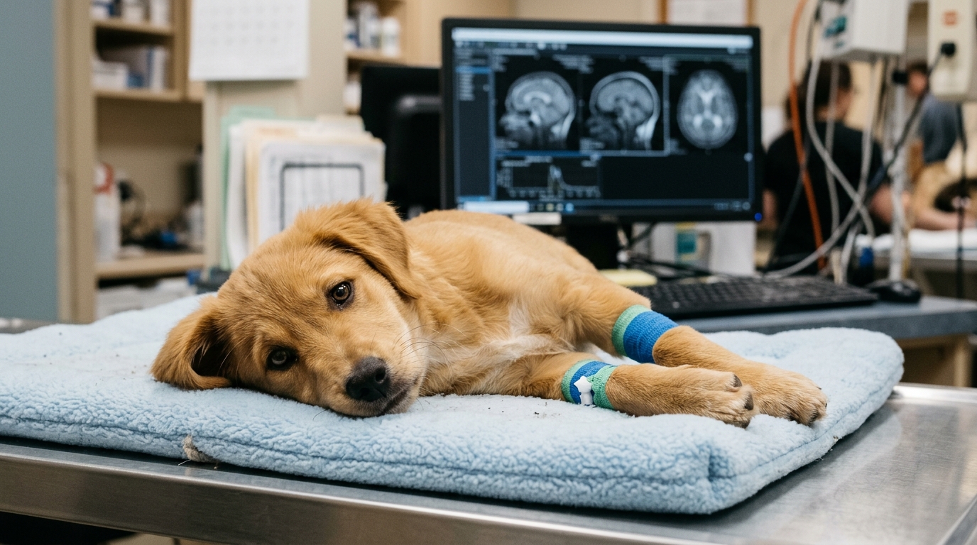
Veterinary clinicians and pathologists have documented an unprecedented case of a cerebral hemangioma presenting with an acute onset of epileptic cluster seizures in a five-month-old female Chow Chow dog. Central nervous system hemangiomas represent benign vascular neoplasms that are exceedingly rare in dogs, typically manifesting in mature adult or geriatric canines with an average risk peak occurring around nine years of age. The acute clinical presentation observed in this juvenile dog was driven primarily by an extensive intralesional and intraventricular hemorrhage secondary to the vascular tumor. Without this dramatic hemorrhagic event, the underlying cerebral mass might have remained clinically silent for a substantial period.
In veterinary neurology, epileptic seizures constitute the most frequently observed clinical presentation, categorized into reactive, idiopathic, or underlying structural etiologies. Published investigations indicate that structural brain disorders account for approximately 45.1 percent of canine seizure presentations, making accurate diagnostic differentiation vital because long-term treatment strategies and clinical prognoses vary substantially between idiopathic epilepsy and structural intracranial lesions. Veterinary patients displaying cluster seizures or persistent interictal neurological deficits carry a significantly increased likelihood of harboring an underlying structural anomaly rather than idiopathic epilepsy. Structural causes encompass inflammatory disease, head trauma, congenital malformations, degenerative conditions, intracranial neoplasia, and acute vascular disorders.
Age distribution plays a pivotal role in formulating clinical differential diagnoses for canine intracranial disease, as brain tumors occur predominantly in older animals. Even the most common primary canine brain tumors, such as astrocytomas and oligodendrogliomas, are exceptionally rare in animals under twelve months of age. Hemangiomas are benign endothelial neoplasms characterized by vascular arrangements closely resembling normal blood vessels. Although canine hemangiomas occur relatively frequently in superficial soft tissues, the dermis, and the hemopoietic system, primary localization within the central nervous system remains distinctly unusual. Prior published reports of canine cerebral hemangiomas have described cases ranging exclusively between thirteen months and nine years of age.
The reported subject was a five-month-old intact female Chow Chow dog that had been imported into Denmark from Greece at fifteen weeks of age. During the final twenty-four hours preceding university presentation, the young dog experienced five distinct generalized epileptic seizures in rapid succession, prompting emergency intervention and the intravenous administration of phenobarbital at an outside veterinary emergency facility. Upon physical admission to the referral teaching hospital, the puppy was non-ambulatory and markedly obtunded, clinical signs indicating a severe intracranial lesion localized within either the forebrain or the brainstem structures.
Physical and neurological evaluations revealed pronounced multifocal abnormalities, including an intermittent right-sided head turn, a complete absence of conscious postural reactions across all four limbs, and preserved spinal withdrawal reflexes. Cranial nerve assessments demonstrated bilaterally decreased menace responses alongside an intermittent positional ventral strabismus affecting the right eye, while the remaining cranial nerves functioned normally. The constellation of acute epileptic cluster seizures, altered consciousness, and postural deficits pointed toward a right-sided forebrain lesion, whereas the intermittent right ventral strabismus suggested additional cerebellar or brainstem involvement. To rule out potential extracranial and systemic metabolic disturbances, clinicians conducted comprehensive paraclinical laboratory screenings. Complete blood counts fell entirely within established canine reference intervals, and serum biochemistry panels revealed only mild elevations in alkaline phosphatase and serum phosphate, both of which represented expected physiological findings in a growing five-month-old juvenile dog.
Further diagnostic evaluations ruled out infectious and toxic etiologies that could mimic primary intracranial pathology in a young imported dog. Point-of-care serological and antigen assays confirmed negative results for Anaplasma species, Borrelia burgdorferi, Dirofilaria immitis, Ehrlichia species, and the canine lungworm Angiostrongylus vasorum. Comprehensive urine toxicological screening panels yielded negative results for widespread illicit and medicinal drugs, including cannabis, opiates, amphetamines, cocaine, methadone, tramadol, buprenorphine, benzodiazepines, fentanyl, ketamine, oxycodone, and nicotine. Because extracranial toxic, infectious, and metabolic factors were definitively eliminated, clinicians prioritized primary structural differential diagnoses, including congenital intracranial malformations, meningoencephalitis of unknown origin, infectious encephalitis, an acute vascular accident, or, considered far less probable due to the patient’s age, primary intracranial neoplasia. Advanced cross-sectional imaging was promptly scheduled to directly examine the patient’s neurocranium.
Magnetic resonance imaging of the head was performed utilizing a high-field 1.5-Tesla Siemens Magnetom Altea system equipped with a specialized head matrix coil. Multiplanar sequences included sagittal and transverse T2-weighted scans, transverse T1-weighted images, transverse fluid-attenuated inversion recovery sequences, transverse T2-weighted gradient echo sequences, diffusion-weighted imaging, and contrast-enhanced T1-weighted views following intravenous gadobutrol administration. The neuroimaging revealed an expansive, well-delineated, multilobulated mass centered in the right frontal lobe, measuring approximately 2.5 centimeters by 1.4 centimeters. The lesion extended anatomically from the olfactory bulb to the rostral aspect of the right lateral ventricle, displaying marked heterogeneous hypo- to hyperintensity on both T1-weighted and T2-weighted images alongside prominent heterogeneous contrast enhancement. Extensive intralesional susceptibility artefacts on gradient echo sequences confirmed abundant internal hemorrhage, with a distinct lateral fluid-fluid level illustrating disparate degradation stages of blood products.
The massive right frontal lobe lesion generated severe secondary intracranial consequences, including substantial leftward midline shift and marked perilesional vasogenic edema tracking along right hemispheric white matter tracts with elevated diffusion. Neuroimaging also documented generalized ventriculomegaly, cervical spinal cord presyrinx formation, and two distinct fluid-filled structures. A 0.9-centimeter extra-axial cyst-like lesion was located in the right cerebellopontine angle, exhibiting cerebrospinal fluid isointensity and complete suppression on fluid-attenuated inversion recovery without contrast enhancement. A second well-defined 1.2-centimeter circular lesion sat within the fourth ventricle, showing hyperintensity on T2-weighted images and partial fluid suppression.
Gross macroscopic examination at necropsy confirmed that pathological lesions were entirely confined to the central nervous system. The leptomeninges overlying the dorsal cerebral hemispheres were markedly thickened, firm, and strikingly erythematous, studded with coalescing, slightly elevated grey plaques that separated easily from the cerebral cortex. Following three weeks of fixation in ten percent neutral buffered formalin, agar-embedded four-millimeter coronal brain sections revealed a firm, well-demarcated, dark hemorrhagic mass compressing the right hemisphere and rupturing directly into the right lateral ventricle, which contained a large organized coagulum. Histological sections stained with hematoxylin and eosin, Masson’s trichrome, periodic acid-Schiff, von Kossa, and Perl’s Prussian blue demonstrated that the neoplasm was composed of numerous irregular, thin-walled vascular channels admixed with massive hemorrhage and focal perivascular fibrosis.
Immunohistochemical evaluations definitively established the cellular identity of both the vascular neoplasm and the concurrent ventricular anomalies. The endothelial cells lining the neoplastic vascular lumens stained strongly for CD31, while smooth muscle actin immunohistochemistry verified the presence of an intact, delicate smooth muscle layer encircling all vascular channels. Numerous hemosiderin-laden macrophages labeled positively with MAC 387 throughout hemorrhagic territories, while adjacent fibrotic meninges displayed subarachnoid collagen deposition with proliferative vessels and occasional microcalcifications. Examination of the caudal brainstem revealed a multilocular cystic structure occupying the cisterna magna, directly communicating with the fourth ventricle through the median aperture. The cyst lining consisted of cuboidal ciliated epithelial cells expressing cytoplasmic vimentin and variable glial fibrillary acidic protein, resting on a dense astroglial meshwork without cytokeratin expression. This immunophenotypic profile confirmed that the cystic lesions represented ependymal diverticula originating from the fourth ventricle.
This landmark report emphasizes that cerebral hemangiomas accompanied by secondary acute hemorrhage must be considered in the clinical differential diagnosis of canine cluster seizures, even in pediatric veterinary patients. While canine intracranial neoplasms are conventionally anticipated to produce insidious signs in older animals, catastrophic hemorrhage from fragile neoplastic blood vessels can produce sudden, apoplectic neurological collapse. Comparative oncology literature indicates that hemangiomas are among the most common infantile neoplasms in humans, where central nervous system lesions are linked to dysregulated vasculogenesis often triggered by local hypoxic stress. Although historical veterinary cases have explored surgical interventions such as lobectomy or experimental viral therapies, and human medicine frequently utilizes beta-blockers like propranolol to inhibit endothelial proliferation, the extreme severity of the acute hemorrhage in this young Chow Chow necessitated immediate humane intervention, providing invaluable diagnostic insights into rare juvenile intracranial vascular tumors.
Subject of Research: Canine cerebral hemangioma and secondary intracranial hemorrhage in a juvenile dog
Article Title: Cerebral hemangioma in a 5-month-old Chow Chow dog displaying seizures of acute onset
Article References: Heidemann, P. L., Pedersen, M. L., Miller, A. D., Allberg, C., & Agerholm, J. S. (2026). Cerebral hemangioma in a 5-month-old Chow Chow dog displaying seizures of acute onset. Acta Veterinaria Scandinavica, 68(1), Article 29. https://doi.org/10.1186/s13028-026-00872-7
Image Credits: AI Generated
DOI: 10.1186/s13028-026-00872-7
Keywords: veterinary neurology, cerebral hemangioma, canine epilepsy, intracranial hemorrhage, magnetic resonance imaging, ependymal diverticulum, neuropathology, Chow Chow, Cerebral, hemangioma, month-old, Chow
News Source: William Thompson. (November 6, 2026). Cerebral Hemangioma Causes Sudden Seizures in a Five-Month-Old Dog. Scienmag.