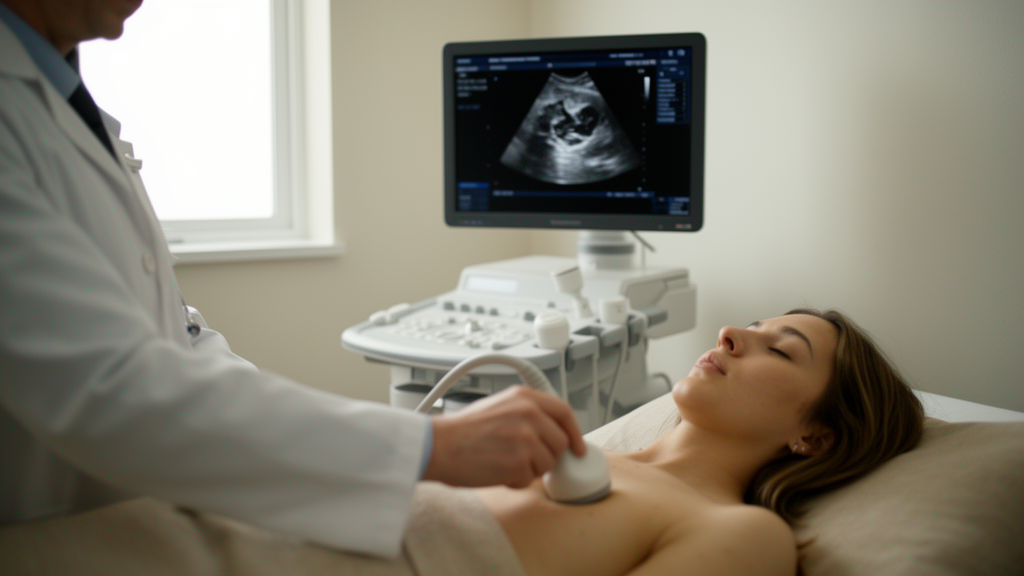
Artificial intelligence built into handheld ultrasound devices can estimate how well a patient’s heart is pumping with impressive accuracy, but a new prospective study finds it still does not match the judgment of trained cardiac sonographers reading the same bedside images. In a single-center comparison at Tufts Medical Center in Boston, AI-assisted focused cardiac ultrasound tracked formal transthoracic echocardiography closely, yet experienced human operators reading the same bedside images by eye came out ahead on nearly every statistical measure, particularly when it came to grading how severe a patient’s left ventricular dysfunction actually was.
The study, published in the journal Cardiovascular Ultrasound, enrolled 222 patients aged 18 or older who were already scheduled for a formal transthoracic echocardiogram, the comprehensive cart-based imaging exam that serves as the reference standard for measuring left ventricular ejection fraction, the percentage of blood the left ventricle ejects with each contraction. Seven participants were excluded because their focused cardiac ultrasound images were not diagnostic, leaving 215 patients in the final analysis. Enrollment ran from December 2020 to March 2022 using a convenience series dictated by hospital workflow, and the data analysis was completed in October 2025.
Each participant underwent two bedside assessments within 48 hours of their formal echocardiogram, both performed on the same EchoNous KOSMOS handheld point-of-care ultrasound device. First, a trained cardiac sonographer acquired apical four-chamber and two-chamber views and gave a global visual estimate of the ejection fraction. Immediately afterward, the same device’s machine-learning algorithm calculated the ejection fraction using the modified Simpson’s biplane method of disks, with all AI-generated tracings and image frames left unmodified so the echocardiographers could not manually correct the measurements. Because the studies were conducted back to back, image quality was congruent between the visual and AI assessments. The formal cart-based echocardiogram, performed by an experienced level-three echocardiographer using the same Simpson’s biplane method, then served as the reference against which both bedside approaches were judged.
The participants had a median age of 63 years, with an interquartile range of 49 to 73, and 83 of them, or 38.6 percent, were female. The median ejection fraction on formal echocardiography was 60 percent. Only 33 participants, or 15.4 percent, had an abnormally low ejection fraction below 50 percent, and just four patients, or 1.9 percent, had severely reduced function below 30 percent, a detail that would prove important for interpreting the study’s most striking results.
When the question was simply whether the ejection fraction was abnormal, both approaches performed well. AI-assisted focused ultrasound achieved an area under the receiver operating characteristic curve of 0.9779, with a 95 percent confidence interval of 0.9591 to 0.9967, a sensitivity of 90.9 percent and a specificity of 95.6 percent for detecting an ejection fraction below 50 percent. The visual assessment by the bedside sonographer was slightly better, with an area under the curve of 0.9961, sensitivity of 90.9 percent and specificity of 97.8 percent. Both methods shared the same sensitivity, and their negative predictive values were identical at 0.98, meaning a normal bedside reading was reassuringly reliable either way. The positive predictive value favored the human reader, at 0.88 compared with 0.79 for the AI.
The gap widened when the analysis turned to continuous agreement and severity grading. The AI measurements showed good agreement with formal echocardiography, with an intraclass correlation coefficient of 0.84, while the visual estimates produced a notably stronger coefficient of 0.97. On average, the AI readings ran 1.16 percentage points below the formal echocardiographic values, with a standard deviation of 5.36 points, whereas the visual estimates averaged just 0.08 points above the reference, with a standard deviation of only 2.60 points. For categorizing dysfunction as normal, mild, moderate or severe, the weighted kappa was 0.89 for the AI and 0.96 for the visual assessment, both excellent but again favoring the trained human eye. Notably, neither method ever misgraded a patient’s dysfunction by more than one category relative to the formal study.
For detecting severely reduced ejection fraction below 30 percent, the visual assessment was perfect, with an area under the curve of 1.0000, sensitivity of 100 percent and specificity of 100 percent. The AI achieved an area under the curve of 0.9923, with sensitivity of 75.0 percent and specificity of 98.6 percent. The authors are careful to flag, however, that these severe-dysfunction statistics rest on a sample of only four patients, a limitation that makes any conclusion about AI performance in this clinically critical subgroup tentative at best.
The findings land at a moment when handheld ultrasound devices with embedded machine learning are spreading rapidly through emergency departments, critical care units and inpatient wards, settings where formal echocardiography is often logistically constrained or unavailable. Focused cardiac ultrasound has already become part of the standard of care for bedside cardiovascular assessment as an adjunct to physical examination, but its effectiveness is limited by operator dependency, and misinterpretation influenced by operator experience remains an ongoing challenge. Prior work cited in the study suggests AI can help narrow the gap between novice and expert users: a multicenter diagnostic study of a deep-learning algorithm found that most clinical parameters assessed by novice nurses agreed with scans by trained sonographers more than 90 percent of the time, and another analysis found novice and experienced operators showed consistent reliability when using AI-assisted ejection fraction measurement, with intraclass correlation coefficients of 0.921 and 0.845 respectively.
Yet the new results make clear that, at least against formally trained cardiac sonographers, current AI has not closed that gap from the other direction. The authors emphasize that their bedside readers had formal training, which does not reflect the full diversity of real-world focused ultrasound users, so the size of the AI-versus-human difference in this study may not generalize to non-cardiologists with less cardiac imaging experience. They also note the single-center design and convenience sampling as limitations, and they call for replication in broader clinical environments, including rural and community settings, before firm conclusions about real-world performance can be drawn.
The practical takeaway is a division of labor rather than a verdict against the technology. AI-assisted focused ultrasound delivered accurate estimates of both the presence and the severity of left ventricular dysfunction, with strong negative predictive value and no misgradings beyond one category, making it a plausible support tool for early learners, trainees and clinicians working where formal imaging is scarce. But for now, the study concludes, AI is best viewed as a complementary tool within the clinician’s broader assessment, not a replacement for expert human interpretation, and continued advances in algorithm training and real-world validation are needed before machine readings can rival the trained bedside eye.
Subject of Research: Diagnostic accuracy of AI-based versus visual assessment of left ventricular ejection fraction using bedside focused cardiac ultrasound
Article Title: The diagnostic accuracy of left ventricular ejection fraction assessment between visual and artificial intelligence-based algorithms on bedside ultrasound
Article References: Kolobaric, N., Beauregard, N., Barbour, W., Prosperi-Porta, G., Parlow, S., Di Santo, P., Abdel-Razek, O., Jung, R., Bradford, W. B., Tsang, M., Pacifici, S., Ramirez, F. D., Huggins, G. S., Bugeja, A., Simard, T., Mathew, R., Hibbert, B., Motazedian, P., & Marbach, J. A. (2026). The diagnostic accuracy of left ventricular ejection fraction assessment between visual and artificial intelligence-based algorithms on bedside ultrasound. Cardiovascular Ultrasound, 24(1), Article 20. https://doi.org/10.1186/s12947-026-00377-3
Image Credits: AI Generated
DOI: 10.1186/s12947-026-00377-3
Keywords: echocardiography, artificial intelligence, point-of-care ultrasound, left ventricular ejection fraction, diagnostic accuracy, focused cardiac ultrasound, machine learning, cardiac imaging, transthoracic echocardiography, heart failure, clinical validation, medical diagnostics
News Source: Ophelia Keating. (October 28, 2026). AI bedside ultrasound reads heart function well but loses to trained sonographers. Scienmag.