


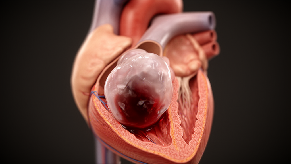
A rare cardiac tumor that looked, on first imaging, like a simple sac of blood has been described in a new case report published in the journal Cardiovascular Ultrasound. The lesion, a balloon-like cystic cardiac myxoma, was found in the left atrium of a 54-year-old woman who had been living with exertional breathlessness, chest tightness and intermittent dizziness for six months. Because the mass appeared as a distended, thick-walled, fluid-filled structure, the team initially suspected a blood-filled cyst rather than a tumor. Only after a sequence of advanced ultrasound techniques, and finally direct observation during surgery, did the true nature of the lesion become clear. The report, authored by Xiaofang Zhong, Shuyu Luo, Lixin Chen and Yingying Liu, was published on 21 September 2026 and documents how a distinctive pattern of blood flow into and out of the mass revealed its identity.
Myxomas are the most common benign tumors of the heart, and according to the report’s introduction they account for approximately 50 percent of all benign primary cardiac tumors. In their typical form they appear as solid masses, and experienced imaging teams usually recognize them before surgery. Occasionally, however, these tumors develop atypical features such as liquefaction, necrosis, hemorrhage or calcification, and these changes can make diagnosis genuinely difficult. Cystic myxomas, in which the tumor takes on a predominantly fluid-filled character, are described as a rare variant with only a few cases reported in the literature. The case now presented is unusual even within that rare group, because the tumor was almost entirely cystic rather than a combination of a superficial cystic shell and a deeper solid core, and because its inflated, balloon-like shape made it resemble a blood-filled cavity rather than a neoplasm.
The clinical story began with a six-month history of symptoms that had recently increased in frequency. When the woman was admitted, her physical examination and electrocardiogram were unremarkable, and laboratory testing, including tumor markers, coagulation tests and immunological assays, also produced no abnormal findings. Despite the mild nature of her symptoms, the imaging work-up that followed uncovered a lesion that the authors judged to carry a high risk of rupture and embolic complications, which is why prompt surgical excision was strongly advised and the patient consented to the operation. The case illustrates how a lesion that produces only modest symptoms can nonetheless pose a serious threat, and how careful imaging characterization is essential to weighing that risk.
Transthoracic echocardiography, the standard first-line ultrasound of the heart, showed a 33 by 29 millimeter balloon-like, thick-walled cystic mass in the left atrium. The mass was attached to the hinge point of the anterior mitral leaflet with a broad base, and it moved with a small-amplitude oscillatory motion that did not obstruct the flow of blood through the mitral valve. Color Doppler imaging added a crucial clue: a narrow pedicle of blood flow was seen entering the cyst from its base. At this stage a blood-filled cyst was the leading suspicion. The finding of a feeding vessel, however, was already a hint that the structure might be more than a passive collection of blood, because a simple blood-filled cyst would not be expected to have its own arterial supply.
Contrast echocardiography was performed next, and it changed the picture substantially. The cystic mass filled with contrast agent, and the cyst cavity showed enhancement that was isointense, meaning equal in intensity, relative to the surrounding myocardium. Two modes were used: a left ventricular opacification mode to better characterize the size, shape and regularity of the mass, and a myocardial contrast echocardiography mode to determine the perfusion pattern of the lesion. A cavity that fills with contrast and enhances like heart muscle is behaving like vascular tissue, not like a static blood pocket. Transesophageal echocardiography was then carried out to obtain more detail, and it revealed a thin flow entering the cyst from its base and another flow exiting from the inferior aspect of the cyst toward the mitral orifice. With an apparent inflow and outflow, and a vascular supply pattern, a benign cardiac tumor was highly suspected.
Surgery provided the definitive confirmation, and it did so in a striking way. During thoracoscopy, the surgeons found a clear, translucent cystic mass that was extensively adherent to the interatrial septum and extended to the mitral valve orifice. After cardioplegic solution was administered into the aortic root to stop the heart for the operation, the solution was seen oozing from the base of the mass, indicating that its vascular supply originated from the aortic root. The influx of the solution was also visualized by intraoperative transesophageal echocardiography. Histopathology then settled the diagnosis: spindle-shaped and stellate tumor cells arranged in cords within a myxoid stroma, accompanied by microvascular proliferation. Immunohistochemistry showed Calretinin positive, CD34 positive, CD31 positive, Factor VIII negative, WT1 negative, CK5/6 negative and partial S-100 positive staining, confirming a benign myxoma.
The outcome for the patient was favorable. She had an uneventful postoperative recovery, and a transthoracic echocardiogram performed before discharge demonstrated complete resolution of the mass with normal mitral valve function. At one-year postoperative follow-up there was no tumor recurrence. The authors note that the mass maintained stable dimensions throughout the cardiac cycle, which they interpret as evidence that arterial inflow continuously equilibrated the pressure inside the cyst with left atrial pressure, with balanced inflow and outflow volumes. This hemodynamic equilibrium helps explain why the lesion held its balloon-like shape on every image rather than expanding or collapsing with each heartbeat.
The report also sets out a proposed mechanism for how a myxoma becomes cystic in the first place. According to the discussion, cystic component formation in a myxoma requires three specific conditions: arterial inflow into the cavity, adequate outflow via drainage orifices, and sufficient integrity of the myxoma membrane. Although the lesion was morphologically similar to previously reported cases, the authors emphasize that their tumor differed markedly in tissue composition, being almost entirely cystic rather than predominantly superficial cystic components over deep solid portions. The vascular supply itself is also of interest. Previous studies have documented myxomas with feeding arteries originating from one or both coronary arteries, a configuration that can potentially cause a coronary artery steal phenomenon. Although coronary angiography was not performed in this case, the color Doppler findings and the observed leakage of cardioplegic solution from the aortic root into the tumor provided evidence that the blood supply arose from the aorta and its branches.
The diagnostic challenge posed by such lesions is considerable, and the authors outline a broad differential diagnosis that includes blood-filled cysts, hemangiomas, angiosarcomas, echinococcal cysts, organized thrombi and metastatic tumors. In their view, the integration of multimodality imaging allows comprehensive evaluation of both morphological and hemodynamic features, and in this case contrast echocardiography and transesophageal echocardiography offered significant insight into the vascular supply pattern of the tumor. They are candid about the limits of ultrasound, however, noting its inherent difficulty in delineating internal cystic components, septations and relationships with adjacent tissue. A stated limitation of the report is the absence of cardiac magnetic resonance imaging, which would have provided additional tissue characterization and facilitated preoperative evaluation. Cardiac magnetic resonance enables clear visualization of internal septations, mural nodules and tumor infiltration into adjacent structures, and late gadolinium enhancement helps differentiate myxomas from thrombi or malignant tumors.
As a single-case report, the study describes one patient’s experience rather than a systematic evaluation of a diagnostic strategy, and no data were generated or analysed in support of the paper beyond the case itself. Its value lies in the detailed documentation of an unusual tumor variant and of the imaging signs, an arterial inflow at the cyst base, an outflow orifice toward the mitral orifice, and contrast enhancement matching the myocardium, that distinguished a benign myxoma from what appeared to be a harmless blood-filled cyst. For clinicians evaluating an apparent cystic mass in the heart, the case is a reminder that a structure with its own inflow and outflow and a demonstrable blood supply deserves consideration as a tumor, with the surgical urgency that classification implies.
Subject of Research: A case report of a rare balloon-like cystic cardiac myxoma in the left atrium diagnosed by multimodality echocardiography and confirmed by surgery and histopathology.
Article Title: A balloon-like cardiac myxoma mimicking a blood-filled cyst
Article References: Zhong, X., Luo, S., Chen, L., & Liu, Y. (2026). A balloon-like cardiac myxoma mimicking a blood-filled cyst. Cardiovascular Ultrasound, 24(1), Article 22. https://doi.org/10.1186/s12947-026-00376-4
Image Credits: AI Generated
DOI: 10.1186/s12947-026-00376-4
Keywords: cardiac myxoma, cystic myxoma, left atrium, transthoracic echocardiography, contrast echocardiography, transesophageal echocardiography, color Doppler, cardioplegia, histopathology, benign cardiac tumor, case report, differential diagnosis
News Source: Ophelia Keating. (October 28, 2026). Rare Balloon-Like Cardiac Myxoma Mimicked a Blood-Filled Cyst. Scienmag.



