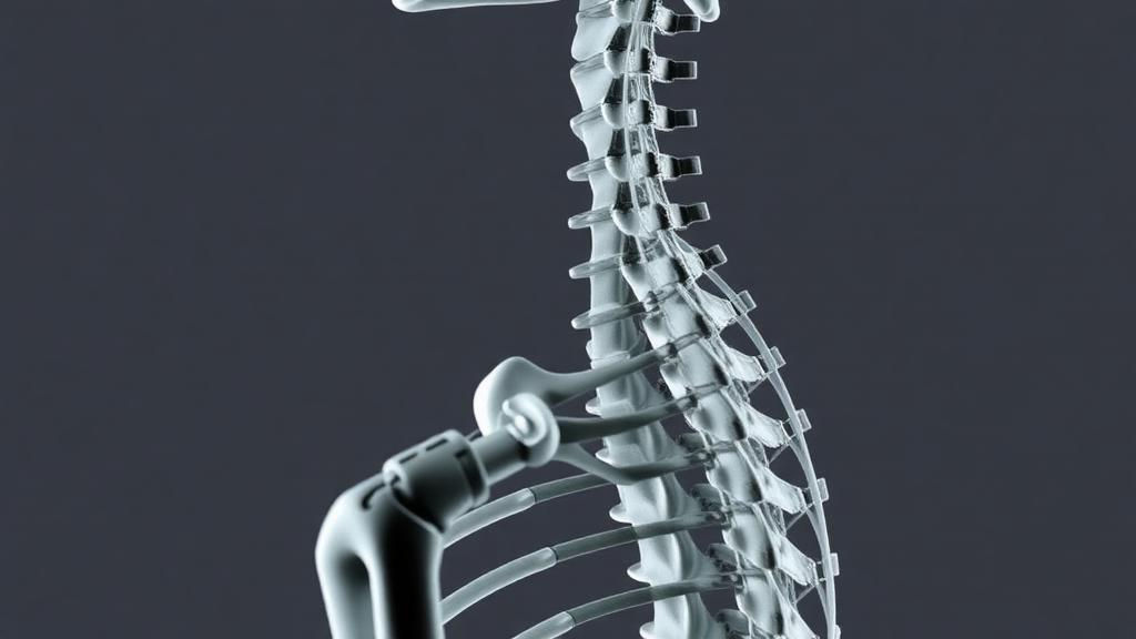
Radiologists may soon have a simpler way to flag broken bones in the spine that patients never knew about, if a new proposal from an invited review in the Egyptian Journal of Radiology and Nuclear Medicine gains traction. The paper, published on 25 October 2026 by Mohamed Ragab Nouh, argues that the biggest obstacle to artificial intelligence–driven osteoporosis screening is not the sophistication of the algorithms but the quality of the labels used to train them. To fix that, the author proposes a deliberately blunt binary classification, called the Skeletal Failure paradigm, which he presents as a hypothesis-generating framework requiring prospective validation rather than an established standard.
The problem the review targets is familiar to anyone who has followed AI in medical imaging. Opportunistic CT screening—extracting bone health information from scans performed for entirely unrelated reasons—has evolved from a diagnostic byproduct into a serious platform for population-level bone assessment. Modern algorithms can segment vertebral bodies, detect compression deformities, and estimate trabecular bone density without calibration phantoms, turning routine abdominal or chest CT scans into screening opportunities. Yet when these models are trained on the traditional semi-quantitative grading scale for vertebral fractures, their performance consistently plateaus.
The culprit, according to the review, is label inconsistency. The long-standing Genant semi-quantitative scale grades deformities as mild, moderate, or severe, and it has served research and clinical trials for decades. But the boundary between normal and mild deformity is inherently subjective. Height loss measurements, the quantitative anchor of the method, vary with endplate selection, vertebral level, and degenerative change. Inter-observer agreement diminishes precisely at that boundary, and nominally gold-standard radiologic labels frequently lack the reliability expected of ground truth. Ambiguous borderline cases introduce systematic label noise, causing algorithms to learn reader-specific tendencies rather than true morphological features. Models trained on high-concordance labels outperform those exposed to heterogeneous annotations, and the review argues that progress may depend less on better architectures than on a more reliable annotation standard.
The proposed solution is a two-pillar definition of Skeletal Failure in which either criterion suffices. The first is unequivocal endplate disruption, visible on CT as buckling, depression, or a cortical step-off. The second is height loss exceeding 20 percent in any dimension—anterior, mid, or posterior—measured from the nearest normal adjacent vertebra, in the absence of a clearly identifiable non-osteoporotic cause such as high-energy trauma or malignancy. The 20 percent threshold corresponds to the lower boundary of Genant grade 1 and is the conventional morphometric cutoff; the author stresses it is intended primarily as a reproducible labeling rule for observer agreement and machine learning annotation, not as a fine prognostic cutoff.
Borderline deformities, such as isolated 15 to 20 percent height loss without endplate abnormality, are deliberately excluded from the binary signal and designated as an indeterminate category. While such findings may carry prognostic significance in population-based studies, their high inter-observer variability and ambiguous clinical actionability make them unsuitable as a standalone triage trigger. Instead, they can be flagged for secondary review or densitometric correlation. The review cites comparative analyses indicating that definitive morphological markers correlate more reliably with biomechanical instability, pain, functional decline, and future fracture risk than borderline deformity gradations do.
Morphology alone, however, is not enough, and the framework pairs it with densitometric context drawn from phantomless CT. Trabecular Hounsfield unit measurements from unfractured vertebrae, obtained without external calibration, predict fracture risk comparably to areal bone mineral density and can forecast subsequent fractures; histological work supports HU as a valid surrogate for trabecular bone quality. A meta-analysis cited in the review reports sensitivity of 0.83 and specificity of 0.74 for CT-based screening. The proposal outlines structured thresholds—above 135 HU as stable, 100 to 135 HU as vulnerable, and at or below 100 HU as high risk—following 2023 ISCD guidance to average two vertebrae such as L1 and L2, with T11 to L2 as a practical alternative when lumbar coverage is incomplete.
Those thresholds come with heavy caveats that the review does not soften. The proposed cutoffs derive from 120 kVp single-scanner data and may not transfer across CT platforms, since tube potential can alter HU values by up to 23 percent. Intravenous contrast elevates trabecular bone density measurements by roughly 9 to 15 milligrams per cubic centimeter on average, an increase not corrected by internal calibration in single-energy CT, though dual-energy CT may offer a correction route that still requires validation. Institutions are advised to derive local thresholds correlated with DXA T-scores or fracture data, and to use the proposed numbers cautiously as guides rather than absolute cutoffs.
The clinical logic of the framework rests on a four-tier triage structure. Category 1 marks Skeletal Failure, Category 2 flags high densitometric risk, Category 3 holds indeterminate or borderline cases, and Category 4 denotes normal or low risk. In the envisioned workflow, AI pre-screening assigns preliminary labels that radiologists confirm or override; indeterminate cases undergo adjudication while negative screens proceed without review, keeping the radiologist in the loop for all positive and indeterminate findings. Structured reports would feed directly into fracture liaison service referral systems or primary care alerts, and the combination of binary morphology and HU-based densitometry would generate machine-readable risk profiles aligned with structured reporting goals.
A substantial portion of the review is devoted to global equity. Access to DXA densitometry, specialist interpretation, and fracture liaison services remains uneven worldwide, and the 1994 WHO T-score definitions, based on Caucasian populations, can underestimate fracture risk in many non-Caucasian groups. The Middle East and North Africa region illustrates the paradox: advanced imaging coexists with marked underdiagnosis. The burden of low bone mineral density in the region nearly doubled over three decades, with 20,371 deaths and 805,959 disability-adjusted life years in 2019 alone, and Saudi Arabia records the highest age-standardized DALY rate at 434.2 per 100,000. A systematic review of 22 Arab countries found vertebral fracture prevalence of 13.3 to 20.2 percent in women, excluding grade 1 deformities, yet most patients were neither tested nor treated. In Kuwait, opportunistic QCT screening of women aged 60 and older found low bone mass in 84.6 percent and vertebral fractures in 21.0 percent, while only 23.9 percent had a prior DXA; in Tunisia, vertebral fractures were present in 26.19 percent of at-risk postmenopausal women; and in Egypt, an estimated 3.36 million women and 2.36 million men have osteoporosis.
The author is candid about the limits. No inter-observer, diagnostic accuracy, clinical outcome, or head-to-head comparison studies of the paradigm have yet been conducted, and key components—including the 20 percent threshold, the HU stratification, and the handling of borderline deformities—lack prospective validation. The framework does not replace established morphometric methods or support longitudinal monitoring, for which the Genant scale remains essential, and its performance depends on adequate CT quality, since artifacts, limited coverage, advanced degeneration, and scoliosis can hinder assessment. Whether removing borderline cases from the training signal actually improves model performance requires direct validation, and expected benefits come with potential unintended consequences such as over-calling benign deformities, unnecessary DXA referral, alert fatigue, and inequitable deployment. The review concludes with a call for prospective validation, comparison of binary versus multi-class approaches, and assessment across diverse populations—positioning the Skeletal Failure paradigm not as a finished clinical tool but as an invitation for the radiology community to test and refine it.
Subject of Research: A proposed binary CT-based classification framework, the Skeletal Failure paradigm, intended to provide reproducible labels for AI-driven opportunistic osteoporosis and vertebral fracture screening.
Article Title: AI-Driven opportunistic osteoporosis screening: the skeletal failure paradigm for trustworthy vertebral fracture triage
Article References: Nouh, M. R. (2026). AI-Driven opportunistic osteoporosis screening: the skeletal failure paradigm for trustworthy vertebral fracture triage. Egyptian Journal of Radiology and Nuclear Medicine, 57(1), Article 227. https://doi.org/10.1186/s43055-026-01895-w
Image Credits: AI Generated
DOI: 10.1186/s43055-026-01895-w
Keywords: osteoporosis, opportunistic CT screening, vertebral fracture, artificial intelligence, machine learning, Hounsfield units, bone mineral density, fracture liaison services, DXA, MENA region, semi-quantitative grading, structured reporting
News Source: Ophelia Keating. (October 28, 2026). Binary ‘Skeletal Failure’ Rule Proposed to Make AI Vertebral Fracture Screening Trustworthy. Scienmag.