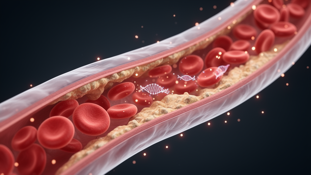
A microRNA measured in a simple blood sample may help identify patients with coronary atherosclerosis, according to a new observational study published in Artery Research. Researchers led by Xingxing Ren, Dongsheng Chen and Zhongcheng Li report that serum levels of miR-20a-5p were distinctly reduced in patients with coronary atherosclerosis compared with individuals whose coronary arteries showed no significant abnormalities, and that the molecule distinguished between the two groups with an area under the ROC curve of 0.841, corresponding to a sensitivity of 81.65 percent and a specificity of 75.25 percent. Beyond its diagnostic signal, laboratory experiments in human vascular smooth muscle cells suggest that reduced miR-20a-5p promotes cellular processes thought to drive atherosclerotic plaque progression, at least in part through the hypoxia-inducible factor HIF1A, which the study identified as a direct target of the microRNA.
Coronary atherosclerosis is a chronic condition characterized by abnormal lipid accumulation and inflammation within the arterial walls, and it remains a major cause of cardiovascular deaths globally. The disease involves multiple interacting processes, including lipid infiltration, inflammatory responses, endothelial dysfunction and thrombus formation. MicroRNAs, short non-coding RNA molecules of roughly 21 to 25 nucleotides, modulate gene expression by interacting with target messenger RNAs, are stable in the blood, and have been identified as potential blood markers in many diseases. Previous studies have linked circulating microRNAs such as miR-126 and miR-130a to coronary atherosclerosis, and other members of the family, including miR-429, miR-30b-5p, miR-125b and miR-146a, have been implicated in endothelial cell apoptosis, smooth muscle cell differentiation, macrophage function and plaque stability. MiR-20a-5p itself had been reported as a mediator of lipid metabolism, inflammation and vascular function in other contexts, but its role in coronary atherosclerosis remained unclear before this investigation.
The study was designed as an observational case-control study conducted at Pingdingshan the Second People’s Hospital between September 2021 and August 2024. It enrolled 109 patients diagnosed with coronary atherosclerosis by coronary angiography and 101 individuals who underwent coronary angiography during the same period but showed no significant abnormalities, defined as stenosis of less than 50 percent in all major coronary branches. Patients with at least one major coronary artery stenosis greater than 50 percent were assigned to the coronary atherosclerosis group. The degree of vascular stenosis was assessed using a combination of physician visual assessment and quantitative coronary angiography, and the angiographic findings were interpreted by two experienced and independent cardiologists under blind conditions. Fasting blood samples were collected from all participants before angiography, and serum was stored at minus 80 degrees Celsius until analysis. The study excluded individuals with infectious diseases or malignant tumors, poorly controlled thyroid dysfunction, prior coronary intervention or surgery, chronic or acute inflammatory and autoimmune diseases, severe renal or hepatic insufficiency, acute cardiovascular events within the previous three months, and a history of bleeding events or contraindications to antiplatelet therapy. The hospital’s ethics committee approved the study, and written informed consent was obtained from all participants. A post hoc power analysis indicated that with the obtained sample sizes, an effect size of 0.5 and an alpha of 0.05, the study achieved a statistical power greater than 89 percent.
The two groups were broadly comparable in age, body mass index, gender, systolic and diastolic blood pressure and high-density lipoprotein cholesterol, with no statistically significant differences in these characteristics. However, the proportions of hypertension and diabetes were significantly higher in the coronary atherosclerosis group, and total cholesterol, triglycerides and low-density lipoprotein cholesterol were all obviously elevated compared with controls. When the variables that differed between the groups were entered into a multivariate logistic regression analysis, hypertension, diabetes and miR-20a-5p each emerged as independent risk factors for coronary atherosclerosis. Hypertension carried an odds ratio of 1.945, diabetes an odds ratio of 2.112, and miR-20a-5p an odds ratio of 0.405, meaning that lower levels of the microRNA were associated with higher odds of the disease after adjustment. Notably, when the researchers used hypertension or diabetes as stratification variables, there was no statistically significant difference in miR-20a-5p expression between the coronary atherosclerosis and control groups, suggesting that the microRNA’s diagnostic signal was not simply a reflection of those comorbidities.
The expression analysis was performed by quantitative reverse transcription PCR on serum samples. Compared with the control group, the average serum miR-20a-5p level in the patient group was distinctly reduced, a difference that reached a P value below 0.001. In the ROC curve analysis, the area under the curve was 0.841 with a 95 percent confidence interval of 0.786 to 0.896, and the optimal cutoff value, determined using the maximum of the Youden index, was 0.765. An area under the curve in this range is generally interpreted as indicating useful discriminatory ability, although the authors emphasize that their cohort was drawn from a single center and that larger, multicenter studies are needed to confirm the diagnostic potential and its generalizability.
To probe the biological function of the microRNA, the team turned to human vascular smooth muscle cells cultured in the laboratory. These cells form the middle layer of arteries and are crucial for maintaining vessel elasticity and contraction. Using a synthetic inhibitor to suppress miR-20a-5p and mimics to raise it, the researchers found that inhibiting the microRNA significantly promoted the proliferation and migration of the cells, whereas overexpression produced the opposite effect, with both outcomes reaching statistical significance at P below 0.001. Additional assays examining other relevant microRNAs, including miR-126, miR-130a, miR-429 and miR-30b-5p, showed no significant effects following treatment with the mimics or inhibitors, which the authors interpret as evidence that the observed phenotypic changes stem specifically from the regulation of miR-20a-5p rather than off-target effects.
The cellular experiments also pointed to mechanisms relevant to plaque formation. Inhibition of miR-20a-5p reduced the expression of SM22α, a marker of the contractile phenotype of vascular smooth muscle cells, while increasing the expression of OPN, a marker of the synthetic phenotype, a shift the authors describe as a key event in the progression of coronary atherosclerosis. At the same time, suppressing the microRNA increased the expression of MMP-2 and MMP-9, matrix metalloproteinases that can degrade the extracellular matrix and are thought to facilitate the migration of smooth muscle cells from the media to the intima, potentially contributing to plaque formation. Raising miR-20a-5p produced the opposite pattern on all of these markers, and measurements at the protein level by western blot confirmed the messenger RNA findings. The authors caution, however, that their study did not directly measure plaque burden or disease progression in living organisms, so these interpretations should be regarded as correlational and hypothesis-generating, requiring further validation in animal models or clinical cohorts.
To identify the molecular target through which miR-20a-5p might act, the researchers searched four bioinformatics databases, miRWalk, miRDB, ENCORI and TargetScan, which collectively yielded 53 candidate genes. Enrichment analyses of these genes highlighted biological processes related to H3-K4 trimethylation and negative regulation of growth, cellular components including the inverter complex and the PI3K complex, and molecular functions such as nuclear receptor activity and lipoprotein receptor activities, while pathway analysis pointed to axon guidance, thyroid hormone signaling and autophagy, all of which have been linked to atherosclerosis in previous work. A protein-protein interaction network constructed from the candidate genes identified HIF1A as the top hub node, with a degree of six, drawing the team’s attention for experimental verification. Dual-luciferase reporter assays then showed that luciferase activity was inversely correlated with miR-20a-5p expression when the wild-type 3′ untranslated region of HIF1A was present, but not when the binding site was mutated, confirming that HIF1A is a direct target of the microRNA. In the patient samples, HIF1A expression was abnormally elevated, and Pearson’s correlation analysis revealed a significant negative correlation between miR-20a-5p and HIF1A, with a correlation coefficient of minus 0.708 and a P value below 0.001.
Finally, the researchers tested whether HIF1A could counteract the effects of the microRNA. When HIF1A was overexpressed in vascular smooth muscle cells with high miR-20a-5p levels, the inhibitory effects of the microRNA on cell proliferation and migration were reversed, and the changes in SM22α, OPN, MMP-2 and MMP-9 were also restored, with the effects reaching statistical significance at the messenger RNA and protein levels. The authors interpret this as evidence that HIF1A overexpression antagonizes the effects of up-regulated miR-20a-5p, clarifying a molecular mechanism by which the microRNA may exert a protective influence through negative regulation of HIF1A. Prior studies have reported that upregulation of HIF1A can promote atherosclerosis and that HIF1A deficiency can alleviate atherosclerosis induced by a high-cholesterol diet, a context that is consistent with the present findings.
The authors are careful to frame their conclusions as preliminary. Beyond the single-center design and the relatively modest sample size, they note that plaque burden was not directly measured, so the relationship between miR-20a-5p and coronary atherosclerosis remains correlational, and that unmeasured confounding mechanisms linking hyperlipidemia to microRNA expression cannot be completely ruled out. They also observe that adjusted lipid levels were not independently associated with disease risk in their regression analysis. Future animal studies, they write, are needed to elucidate the causal role of miR-20a-5p, and multicenter cohorts are required before any clinical translation of the diagnostic or therapeutic implications. Within those limits, the study adds a candidate blood-based marker to the growing list of microRNAs under investigation in cardiovascular disease and proposes a specific regulatory axis, miR-20a-5p and HIF1A, that may shape how vascular smooth muscle cells behave in the walls of diseased coronary arteries.
Subject of Research: Diagnostic value and regulatory role of the microRNA miR-20a-5p in coronary atherosclerosis
Article Title: Diagnostic Value of miR-20a-5p and its Role in Modulating Coronary Atherosclerosis Progression via HIF1A
Article References: Ren, X., Chen, D., & Li, Z. (2026). Diagnostic Value of miR-20a-5p and its Role in Modulating Coronary Atherosclerosis Progression via HIF1A. Artery Research, 32(1), Article 31. https://doi.org/10.1007/s44200-026-00128-9
Image Credits: AI Generated
DOI: 10.1007/s44200-026-00128-9
Keywords: coronary atherosclerosis, miR-20a-5p, HIF1A, biomarker, vascular smooth muscle cells, ROC analysis, phenotypic transition, MMP-2, MMP-9, case-control study, microRNA, diagnostic marker
News Source: Ophelia Keating. (October 28, 2026). Blood MicroRNA Shows Diagnostic Promise and Regulatory Role in Coronary Atherosclerosis. Scienmag.