A routine X-ray taken after a young athlete fell on his outstretched hand has delivered one of the rarest diagnoses in skeletal medicine: melorheostosis, an extraordinary bone disorder so scarce that it affects an estimated 0.9 people per million, striking the small bones of the wrist and hand in fewer than one in ten known cases. The case, published in Clinical Case Reports, describes how a 21-year-old right-hand-dominant man was found to have dense, wax-like bone lesions flowing across his second metacarpal, lunate, and triquetrum—findings so characteristic that clinicians were able to secure the diagnosis entirely without a biopsy, relying instead on a carefully orchestrated combination of radiographs, computed tomography, and magnetic resonance imaging.
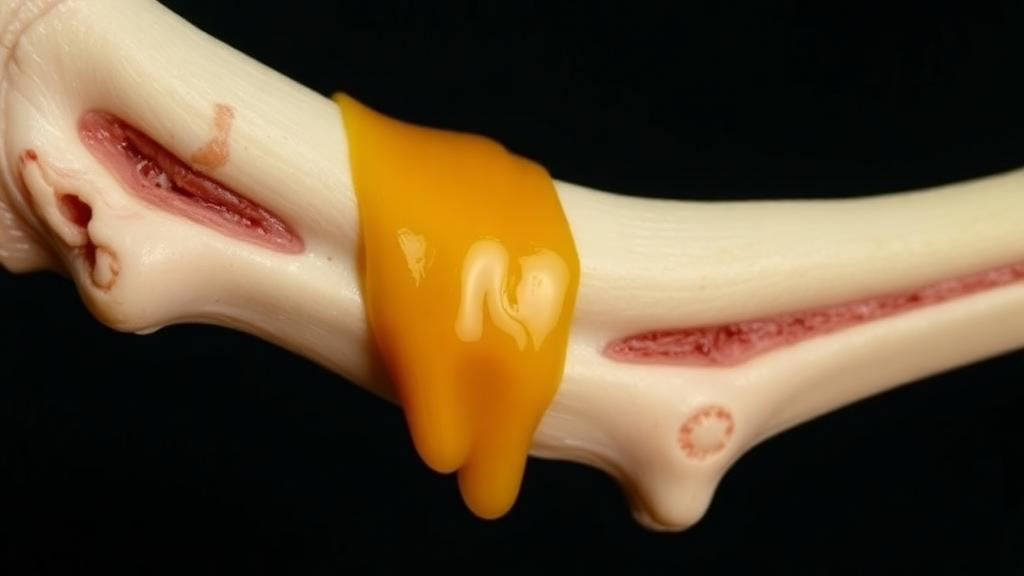
Melorheostosis, also known as Léri-Joanny syndrome, has fascinated physicians since it was first described in 1922. Its nickname in radiology circles, the dripping candle wax sign, comes from the way dense bone appears to ooze along the outer contours of affected bones, as if molten wax had been poured over them and hardened in place. The condition is a non-hereditary, congenital sclerotic bone dysplasia, meaning that bone tissue becomes abnormally thickened and dense. Although the disease was long understood only through its appearance on imaging, recent molecular work has finally revealed its biological roots: somatic activating mutations in the MAP2K1 gene, which encodes the MEK1 protein, drive constitutive activation of the MAPK/ERK signaling pathway. That pathway, when stuck in the on position, pushes osteoblasts—the cells responsible for building bone—into aberrant, uncontrolled activity, producing the dense sclerotic masses that define the disease.
The story of this particular case began on what clinicians call day zero, when the patient fell onto his left hand during recreational sports. He reported only mild wrist discomfort, and his initial examination at an orthopedics clinic the following day revealed localized tenderness over the thenar eminence, the fleshy pad at the base of the thumb. There was no redness, no deformity, no sensory deficit, and his wrist and finger joints moved through a full, painless range of motion. Radiographs ordered simply to rule out a fracture instead unveiled something unexpected: irregular, linear, hyperdense sclerosis involving the second metacarpal, the lunate and triquetrum of the wrist, and the distal ends of the radius and ulna.
Those first images displayed the hallmark ivory-like density and flowing pattern along the bone contours that immediately suggested the dripping candle wax appearance. Critically, there was no periosteal reaction—the aggressive bone-forming response often seen with tumors or infection—and no soft tissue swelling. To characterize the lesions further, the team turned to computed tomography, whose axial and coronal reformats provided exquisite osseous detail. The CT scans confirmed irregular endosteal and intracortical hyperdensity, meaning the dense bone was forming inside the medullary cavity and within the cortex itself. Cortical thickening and partial obliteration of the medullary cavity were evident, and the sclerotic areas blended seamlessly with the surrounding cortex, recreating the melted wax appearance in three dimensions while sparing the adjacent joints.
Magnetic resonance imaging then supplied the decisive piece of the diagnostic puzzle. On both T1-weighted and fat-suppressed T2-weighted sequences, the lesions showed homogeneous, markedly low signal intensity, matching that of cortical bone—exactly what dense, compact sclerotic bone should look like. Far more important was what the MRI did not show: there was no bone marrow edema, no soft tissue mass, and no joint effusion, and the surrounding marrow signal was entirely normal. In bone imaging, the absence of these features is often more informative than their presence, because marrow edema and soft tissue masses are the fingerprints of aggressive processes such as osteosarcoma or osteomyelitis.
Laboratory findings reinforced the benign interpretation. The patient’s complete blood count, C-reactive protein of 2 mg/L, and erythrocyte sedimentation rate of 3 mm/h all fell comfortably within reference ranges, as did serum alkaline phosphatase at 51 U/L and calcium at 9.0 mg/dL. Taken together, the normal inflammatory markers and metabolic panel argued strongly against infection or a metabolic bone disorder. With the classic radiographic wax-flow pattern, CT confirmation of cortical sclerosis, and a conspicuously quiet MRI, the clinicians established a definitive diagnosis of melorheostosis and classified it within the two major frameworks used in the literature. Under Freyschmidt’s radiological classification, the case fits the classic candle-wax subtype, and according to the system proposed by Yu and colleagues, it represents the endosteal type, defined by the direction in which the lesion spreads within the bone.
The rarity of this anatomical distribution deserves emphasis. In Freyschmidt’s review of 23 cases, only five involved the upper extremity, roughly 22 percent, and documented involvement of the lunate and triquetrum—two of the eight small carpal bones of the wrist—is exceedingly scarce. The authors contrasted their case with a recent pediatric report by Na and colleagues, in which a 9-year-old boy presented with multifocal melorheostosis of the entire right upper limb, spanning the scapular coracoid process, humeral head, distal humerus, ulnar head, wrist, and metacarpals. That child exhibited the same pathognomonic melting wax pattern and the same endosteal subtype, but required a biopsy for definitive diagnosis, with histopathology revealing irregular endosteal bone proliferation composed of densely arranged lamellar bone and enlarged, distorted Haversian canals. The adult patient in the new report, by contrast, avoided any invasive procedure altogether, illustrating how multimodal imaging can spare cooperative adult patients the risks and discomfort of surgical sampling.
The comparison also highlights the remarkable phenotypic spectrum of the disease. One patient was a child with multifocal disease throughout an entire limb; the other was an adult with localized involvement confined to the carpometacarpal region. Both remained asymptomatic over their respective follow-up periods—six months in the pediatric case and two years in the present one—reinforcing a consistent management principle: asymptomatic melorheostosis requires monitoring only. The authors acknowledge one limitation of their report, namely that no repeat imaging was performed during follow-up because the absence of symptoms did not justify additional radiation exposure. Still, a sustained, completely asymptomatic clinical course over two years strongly supports the interpretation of a benign, quiescent process.
The molecular dimension of the disease adds a forward-looking note to the discussion. Because MAP2K1 mutations arise somatically after fertilization, they produce post-zygotic mosaicism, which elegantly explains why the disease tends to affect localized, often linear distributions along a single limb rather than the entire skeleton. Genetic testing was not performed in this asymptomatic patient, consistent with current clinical practice in which diagnosis remains imaging-based, but the authors stress that recognizing these molecular underpinnings is crucial. Beyond explaining the disease’s localized nature, the identification of a specific druggable pathway—the MEK-ERK cascade—opens potential avenues for targeted therapies in symptomatic cases, a prospect that was unimaginable when the condition was first described a century ago.
Differentiating melorheostosis from its mimics remains the central diagnostic challenge, particularly when lesions are discovered incidentally in the wrist. The differential diagnosis in this case included osteosarcoma, chronic osteomyelitis, and osteopoikilosis, a related sclerotic bone dysplasia often associated with LEMD3 mutations that produces multiple small, round sclerotic foci but lacks the linear, flowing morphology of melorheostosis. Osteosarcoma, the most dangerous mimic, typically shows heterogeneous signal intensity on MRI, a soft tissue mass, and aggressive periosteal reaction—features conspicuously absent here. The authors caution that the absence of marrow edema alone cannot entirely exclude a low-grade malignancy, which is precisely why the diagnosis rested on the overall clinical and radiological constellation rather than any single imaging feature. Treatment, when needed at all, is reserved for complications such as nerve entrapment, progressive deformity, or intractable pain; this patient received only reassurance, education, and periodic clinical follow-up, with instructions to return if pain or functional limitation ever develops. At his two-year check, he remained entirely symptom-free—a quiet outcome that, for a disease once guaranteed to trigger invasive work-ups, represents a genuine victory for modern diagnostic imaging.
Subject of Research: Non-invasive multimodal imaging diagnosis of melorheostosis involving the carpal and metacarpal bones
Article Title: Melorheostosis Involving the Carpal and Metacarpal Bones: Diagnostic Insights From Multimodal Imaging
Article References: Zhang, Z., Deng, Z., Yang, W., Zou, A., Leng, P., & Peng, Y. (2026). Melorheostosis Involving the Carpal and Metacarpal Bones: Diagnostic Insights From Multimodal Imaging. Clinical Case Reports, 14(10), Article e73526. https://doi.org/10.1002/ccr3.73526
Image Credits: AI Generated
DOI: 10.1002/ccr3.73526
Keywords: melorheostosis, bone dysplasia, radiology, MRI, computed tomography, MAP2K1, MAPK/ERK pathway, wrist, candle wax sign, sclerotic bone lesions, differential diagnosis, case report
News Source: Ophelia Keating. (October 6, 2026). Rare Dripping Candle Wax Bone Disease Diagnosed Without a Single Biopsy. Scienmag.