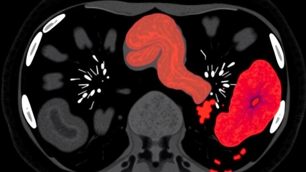
Cholangiocarcinoma, a malignant tumor arising from the bile ducts, is one of the most treacherous diagnoses in hepatobiliary medicine. It carries a poor prognosis, and its imaging appearance on contrast-enhanced computed tomography frequently overlaps with that of benign conditions affecting the gallbladder and bile ducts, such as inflammation, stones, and strictures. The diagnostic ambiguity is particularly severe for perihilar disease, where tumors sit at the confluence of the right and left hepatic ducts and can masquerade as benign scarring or inflammatory change. A misdiagnosis in either direction carries serious consequences: a missed cancer can delay curative surgery until the disease has advanced beyond resectability, while an unnecessary operation for a benign condition exposes patients to major hepatic resection with substantial morbidity. Now, a team of researchers in South Korea reports a fully automated deep-learning system that reads multiphase CT scans in a fundamentally new way, using a technique called supervised contrastive learning to teach an algorithm what distinguishes cancerous tissue from benign disease across the entire three-dimensional biliary tree.
The study, published in BMC Medical Imaging, was conducted by Hee Chang Lee, Woo Hyun Kim, Giltae Song, Jin Kyu Gahm, and colleagues at Pusan National University and Pusan National University Hospital. Their goal was to overcome two persistent limitations of prior CT-based artificial intelligence approaches to cholangiocarcinoma. First, most previous studies relied on single-phase images, typically the portal venous phase, discarding the rich temporal information contained in the arterial, portal venous, and delayed phases of a contrast-enhanced examination. Second, earlier models generally depended on expert-defined two-dimensional regions of interest, in which a radiologist manually outlines the suspicious lesion. That manual step not only consumes specialist time but also constrains the algorithm to a flat slice of anatomy, preventing it from learning the true three-dimensional geometry of the biliary system, which winds through the liver and spans both intrahepatic and extrahepatic ducts.
The technical architecture of the new system unfolds in two stages. In the first stage, the researchers applied automated segmentation networks to identify the liver and gallbladder on each CT scan, and from these structures they extracted three-dimensional hepatobiliary patches, volumetric blocks of image data that cover the intrahepatic and extrahepatic bile ducts without any human outlining. This automated patch extraction is what makes the pipeline fully self-contained: given a raw multiphase CT, the system can localize the relevant anatomy and prepare the model inputs without radiologist interaction. The segmentation step was validated with standard overlap and boundary metrics, including the Dice similarity coefficient, Hausdorff distance, and normalized surface dice, ensuring that the extracted volumes reliably encompassed the biliary structures of interest.
The second and most distinctive stage is the supervised contrastive pre-training. In this paradigm, the network is not initially taught to output a diagnosis. Instead, it learns an internal representation of the images by comparing pairs of examples. Crucially, the researchers constructed positive pairs from the same patient: the arterial-phase patch and the delayed-phase patch of one individual were presented to the model as two views of the same underlying case. The contrastive objective pulls together the learned feature embeddings of these paired patches while pushing apart the embeddings of patches from different patients, and the supervision signal ensures that examples sharing the same diagnostic label cluster together in the embedding space. By training across phases in this way, the network is forced to discover features that are consistent within a patient yet discriminative between disease states, effectively learning how cancer and benign disease manifest across the dynamic enhancement patterns of the liver and bile ducts rather than in a single static snapshot.
After this pre-training, the model was fine-tuned as a classifier on portal venous phase patches, the phase in which many hepatobiliary lesions are conventionally evaluated. The retrospective evaluation enrolled 427 patients: 129 with cholangiocarcinoma, 179 with benign gallbladder disease, and 119 with benign bile duct disease. Performance was assessed with stratified five-fold cross-validation, a rigorous scheme in which the data are divided into five partitions and the model is trained and tested five separate times so that every patient appears in a test set exactly once. The researchers reported sensitivity, specificity, balanced accuracy, and the area under the receiver operating characteristic curve, summarizing results across the five folds to capture the stability of the system rather than a single lucky run.
The results demonstrate meaningful diagnostic power. For the task of distinguishing cholangiocarcinoma from benign gallbladder disease, the model achieved a balanced accuracy of 82.43 percent with an area under the curve of 89.79 percent. Differentiating cancer from benign bile duct disease proved harder, as expected given the closer imaging overlap, but the system still reached a balanced accuracy of 69.90 percent and an area under the curve of 78.26 percent. In both tasks, the proposed phase-aware contrastive model achieved a significantly higher pooled area under the curve than every single-phase model it was compared against, with all differences passing Holm-adjusted statistical significance thresholds. It also consistently exceeded a multiphase early-fusion baseline, which simply concatenates the phases as input channels, across five repeated cross-validation runs, although that particular difference did not reach statistical significance in the primary analysis. The pattern of results suggests that the benefit comes not merely from seeing more phases but from learning how the phases relate to one another.
To understand what the network was actually looking at, the team applied Grad-CAM, a visualization technique that highlights the image regions most influential in a model’s prediction. The resulting maps localized prediction-relevant features to periductal regions and intrahepatic parenchymal areas, precisely the zones where cholangiocarcinoma produces its characteristic changes, such as ductal wall thickening, periductal infiltration, and altered parenchymal enhancement. This anatomical plausibility matters for clinical trust: an algorithm whose attention falls on irrelevant structures would be difficult to deploy, whereas one that concentrates on the ducts and surrounding liver tissue behaves in a way that radiologists can inspect and reason about. Interpretability tools of this kind are increasingly regarded as essential for moving medical artificial intelligence from benchmark performance to bedside utility.
The clinical implications are considerable. Because the pipeline is fully automated, it could in principle run in the background of a radiology workflow, flagging CT studies whose features resemble cholangiocarcinoma and prompting closer review, endoscopic ultrasound, or magnetic resonance imaging follow-up. The authors position the system as a complementary decision-support tool rather than a replacement for expert interpretation, with the aim of improving diagnostic consistency, particularly for the challenging perihilar cases where human readers most often disagree. In an era when imaging volumes are rising and subspecialty expertise is unevenly distributed, a second reader that never tires and always applies the same learned criteria could reduce the variability that currently plagues biliary tumor diagnosis. The researchers also note the potential for integration into clinical workflows, a step that would require prospective validation on external datasets and regulatory evaluation.
The study was approved by the Institutional Review Board of Pusan National University Hospital and conducted in accordance with the Declaration of Helsinki, with written informed consent obtained from all participants. The work was funded by the Institute of Information and Communications Technology Planning and Evaluation through grants from the Korean Ministry of Science and ICT, by the National R&D Program for Cancer Control of the Ministry of Health and Welfare, by a Biomedical Research Institute grant from Pusan National University Hospital, and by the Seegene Medical Foundation. The authors declared no competing interests, and the article is published open access under a Creative Commons Attribution 4.0 license.
Looking forward, the approach illustrates a broader trend in medical imaging artificial intelligence: the shift from hand-crafted regions of interest and single-snapshot analysis toward self-supervised and contrastively learned representations that exploit the full richness of modern imaging protocols. Multiphase CT is already acquired routinely in hepatobiliary workups, yet much of its temporal information goes unused by conventional pipelines. By treating different contrast phases of the same patient as naturally paired views, supervised contrastive learning turns routine clinical data into a training signal that requires no additional annotation burden. If validated prospectively, this strategy could extend beyond cholangiocarcinoma to other cancers whose diagnosis hinges on subtle, phase-dependent enhancement patterns, offering a template for building diagnostic systems that see the way contrast itself reveals disease.
Subject of Research: Deep-learning differentiation of cholangiocarcinoma from benign hepatobiliary disease using multiphase CT and supervised contrastive learning
Article Title: Multiphase CT-based differentiation of cholangiocarcinoma using supervised contrastive learning
Article References: Lee, H. C., Kim, W. H., Park, S. H., Lee, H. J., Lee, J. H., Han, S. Y., Song, G., Kim, D. U., & Gahm, J. K. (2026). Multiphase CT-based differentiation of cholangiocarcinoma using supervised contrastive learning. BMC Medical Imaging. https://doi.org/10.1186/s12880-026-02835-w
Image Credits: AI Generated
DOI: 10.1186/s12880-026-02835-w
Keywords: cholangiocarcinoma, bile duct cancer, multiphase CT, supervised contrastive learning, deep learning, medical imaging, computer-aided diagnosis, biliary tract disease, radiology, machine learning, Grad-CAM, Pusan National University
News Source: Nathaniel Bowman. (October 6, 2026). AI Learns to Spot Bile Duct Cancer on Multiphase CT Scans. Scienmag.